Copyright
©The Author(s) 2019.
World J Clin Cases. Oct 26, 2019; 7(20): 3358-3363
Published online Oct 26, 2019. doi: 10.12998/wjcc.v7.i20.3358
Published online Oct 26, 2019. doi: 10.12998/wjcc.v7.i20.3358
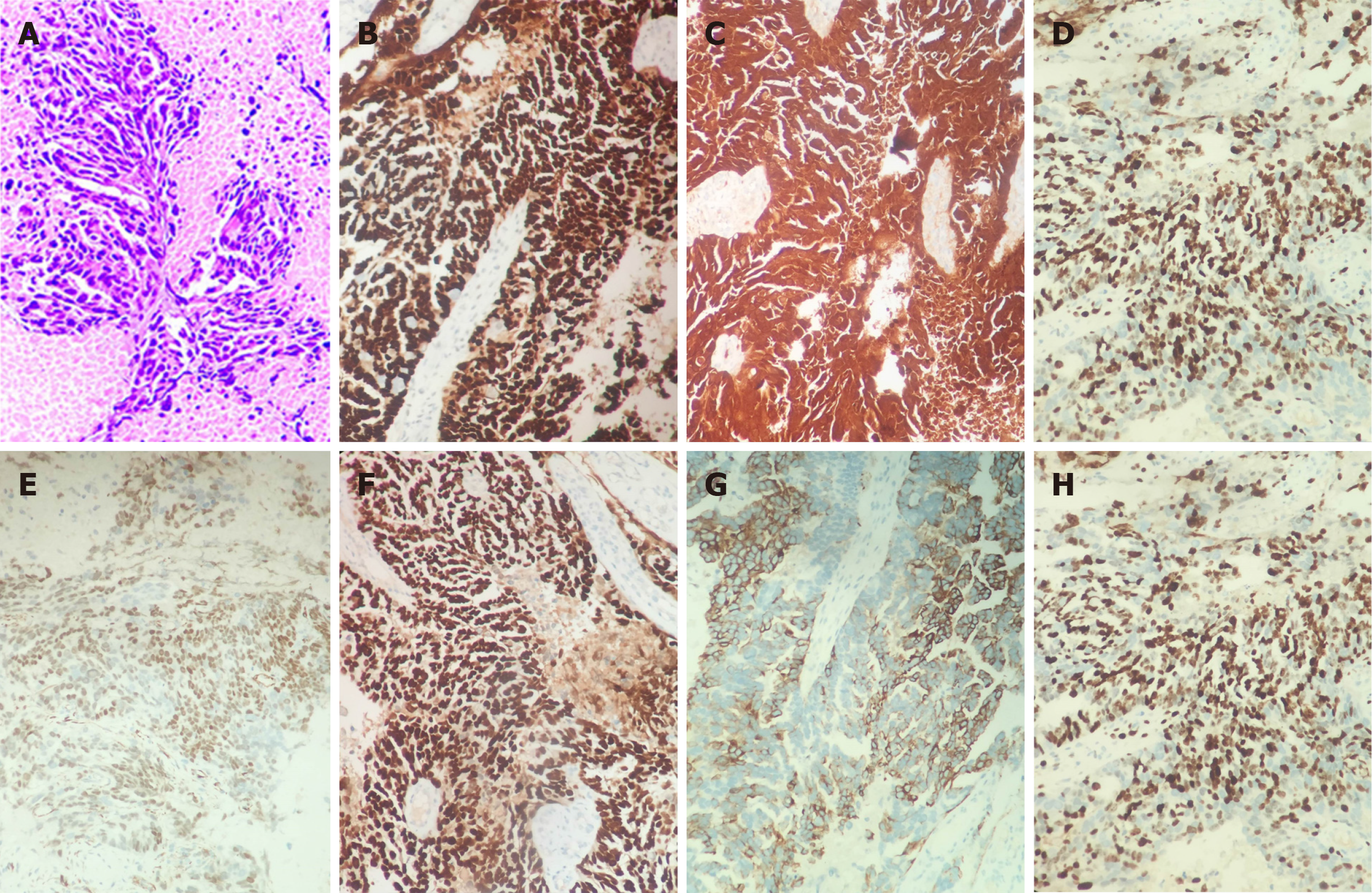
Figure 3 Morphology of metastatic specimens (Hematoxylin and eosin, × 100) and immumohistochemical staining of cancer cells in metastatic lesions (× 200).
A: Dark purple shows adenoid cancer cells; B: Positive staining of umbilical metastasis with anti-P53 antibody; C: Positive staining of umbilical metastasis with anti-P16 antibody; D: Positive staining of umbilical metastasis with anti-ER antibody; E: Positive staining of umbilical metastasis with anti-WT-1 antibody; F: Positive staining of umbilical metastasis with anti-PAX-8 antibody; G: Positive staining of umbilical metastasis with anti-CK7 antibody; H: Positive staining of umbilical metastasis with anti-Ki-67 antibody.
- Citation: Li Y, Guo P, Wang B, Jia YT. Sister Mary Joseph’s nodule in endometrial carcinoma: A case report. World J Clin Cases 2019; 7(20): 3358-3363
- URL: https://www.wjgnet.com/2307-8960/full/v7/i20/3358.htm
- DOI: https://dx.doi.org/10.12998/wjcc.v7.i20.3358


