Copyright
©The Author(s) 2019.
World J Clin Cases. Oct 26, 2019; 7(20): 3289-3295
Published online Oct 26, 2019. doi: 10.12998/wjcc.v7.i20.3289
Published online Oct 26, 2019. doi: 10.12998/wjcc.v7.i20.3289
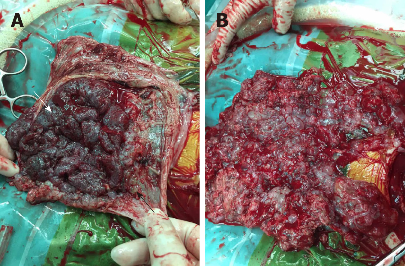
Figure 4 Histopathology of the hydatidiform mole.
Chorionic villi of varying sizes and shapes and focal trophoblastic hyperplasia (hematoxylin and eosin staining; magnification, ×40). A and B: Focal hydropic villi with an irregular scalloped outline and focal trophoblastic hyperplasia between hydrops.
- Citation: Zhang RQ, Zhang JR, Li SD. Termination of a partial hydatidiform mole and coexisting fetus: A case report. World J Clin Cases 2019; 7(20): 3289-3295
- URL: https://www.wjgnet.com/2307-8960/full/v7/i20/3289.htm
- DOI: https://dx.doi.org/10.12998/wjcc.v7.i20.3289


