Copyright
©The Author(s) 2019.
World J Clin Cases. Oct 26, 2019; 7(20): 3259-3265
Published online Oct 26, 2019. doi: 10.12998/wjcc.v7.i20.3259
Published online Oct 26, 2019. doi: 10.12998/wjcc.v7.i20.3259
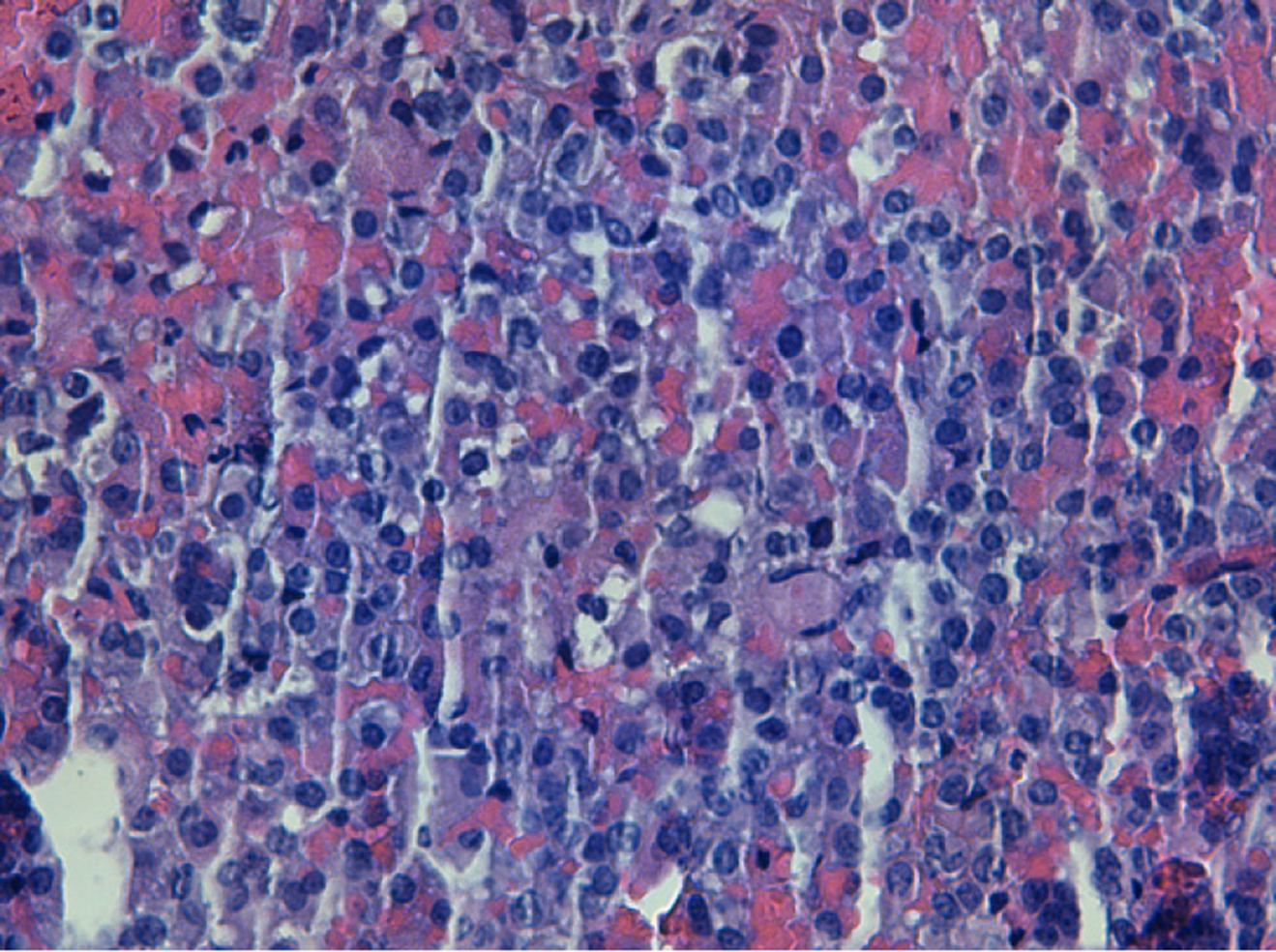
Figure 2 Microscopic findings of the pituitary adenoma.
It shows proliferation of ovoid cells, monotonous, with irregular chromatin, without showing cytological atypia or mitosis figures (× 200).
- Citation: Triviño V, Fidalgo O, Juane A, Pombo J, Cordido F. Gonadotrophin-releasing hormone agonist-induced pituitary adenoma apoplexy and casual finding of a parathyroid carcinoma: A case report and review of literature. World J Clin Cases 2019; 7(20): 3259-3265
- URL: https://www.wjgnet.com/2307-8960/full/v7/i20/3259.htm
- DOI: https://dx.doi.org/10.12998/wjcc.v7.i20.3259


