Copyright
©The Author(s) 2019.
World J Clin Cases. Oct 26, 2019; 7(20): 3194-3201
Published online Oct 26, 2019. doi: 10.12998/wjcc.v7.i20.3194
Published online Oct 26, 2019. doi: 10.12998/wjcc.v7.i20.3194
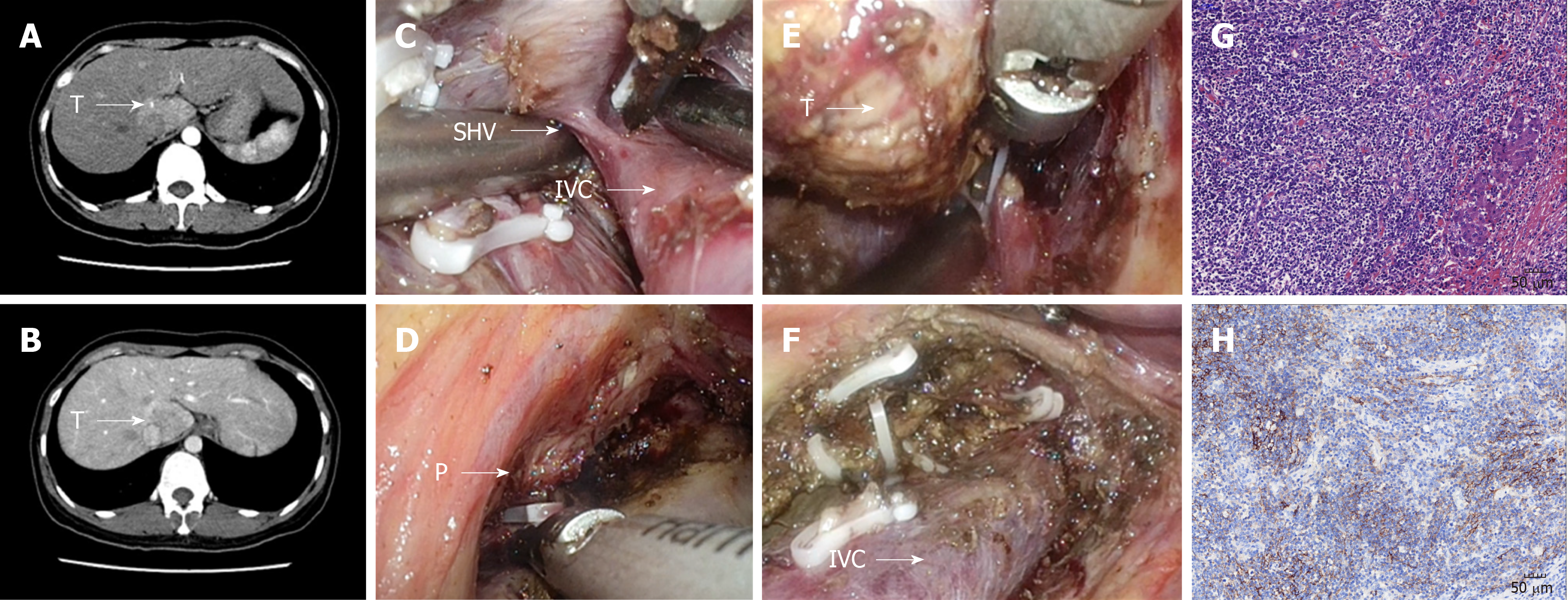
Figure 1 Combined approach to laparoscopic caudate inflammatory pseudotumor-like follicular dendritic cell sarcoma lobectomy.
A and B: The tumor was located at the junction of the Spiegel’s lobe and the paracaval portion; C: The short hepatic vein was dissected; D: The feeding portal pedicle of the caudate lobe (arrow P); E: Isolation of the caudate lobe from right side; F: Surgical area after the tumor was resected; G: Microscopic appearance of caudate inflammatory pseudotumor-like follicular dendritic cell sarcoma (×200); H: Positive CD21 staining by immunohistochemistry (×200).
- Citation: Li Y, Zeng KN, Ruan DY, Yao J, Yang Y, Chen GH, Wang GS. Feasibility of laparoscopic isolated caudate lobe resection for rare hepatic mesenchymal neoplasms. World J Clin Cases 2019; 7(20): 3194-3201
- URL: https://www.wjgnet.com/2307-8960/full/v7/i20/3194.htm
- DOI: https://dx.doi.org/10.12998/wjcc.v7.i20.3194


