Copyright
©The Author(s) 2019.
World J Clin Cases. Jan 26, 2019; 7(2): 242-252
Published online Jan 26, 2019. doi: 10.12998/wjcc.v7.i2.242
Published online Jan 26, 2019. doi: 10.12998/wjcc.v7.i2.242
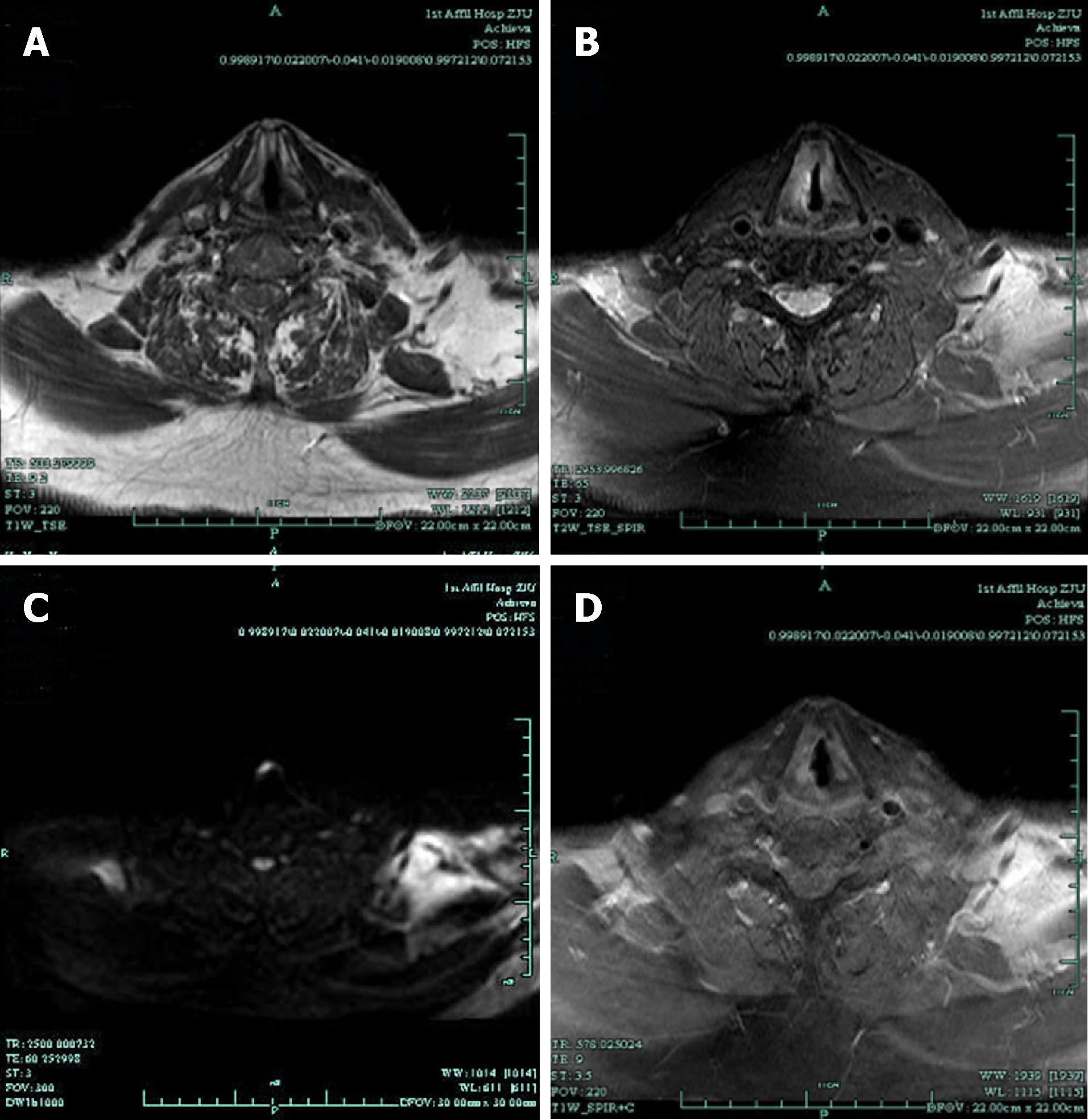
Figure 2 Magnetic resonance imaging images.
Magnetic resonance imaging (MRI) with contrast revealed that the right vocal cord was thicker than the left. A: T1-weighted imaging was isointense; B: T2-weighted imaging was hyperintense; C: Diffusion-weighted imaging was hyperintense; D: Contrast-enhanced T1-weighted MR image showed obvious enhancement.
- Citation: Yu Q, Chen YL, Zhou SH, Chen Z, Bao YY, Yang HJ, Yao HT, Ruan LX. Collision carcinoma of squamous cell carcinoma and small cell neuroendocrine carcinoma of the larynx: A case report and review of the literature. World J Clin Cases 2019; 7(2): 242-252
- URL: https://www.wjgnet.com/2307-8960/full/v7/i2/242.htm
- DOI: https://dx.doi.org/10.12998/wjcc.v7.i2.242


