Copyright
©The Author(s) 2019.
World J Clin Cases. Jan 26, 2019; 7(2): 215-220
Published online Jan 26, 2019. doi: 10.12998/wjcc.v7.i2.215
Published online Jan 26, 2019. doi: 10.12998/wjcc.v7.i2.215
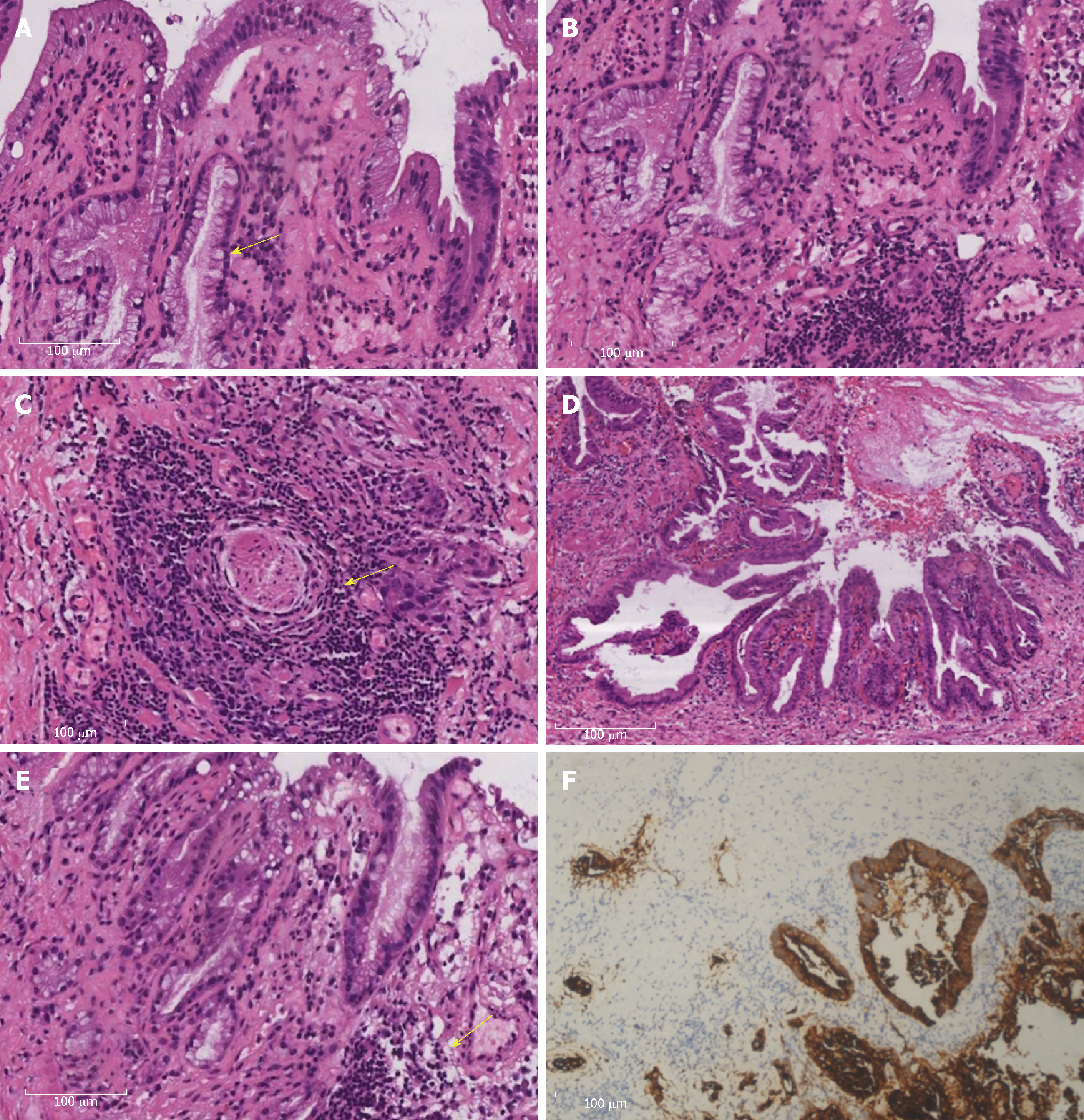
Figure 2 Histopathological appearances of the concomitant tumors in the extrahepatic bile duct.
A: At higher magnification, the area of adenocarcinoma (AC) showed gland-like differentiation (arrow) and mucin production (HE, × 20); B: A transition area between AC and squamous cell carcinoma (SCC) was visible (HE, × 20); C: The area of SCC showed individual cell keratinization and the presence of inter-cellular bridges (HE, × 20). Note the presence of keratin pearls (arrow); D: Low magnification of papillary cystadenocarcinoma (HE, × 4); E: Higher magnification of moderately differentiated cystadenocarcinoma with a glandular pattern, and mild to moderate nuclear atypia (arrow), without mesenchymal stroma (HE, × 20); F: Immunostaining for Cytokeratin 7 (CK7) in the AC component of the common bile duct. HE: Hematoxylin and eosin.
- Citation: Lu BJ, Cao XD, Yuan N, Liu NN, Azami NL, Sun MY. Concomitant adenosquamous carcinoma and cystadenocarcinoma of the extrahepatic bile duct: A case report. World J Clin Cases 2019; 7(2): 215-220
- URL: https://www.wjgnet.com/2307-8960/full/v7/i2/215.htm
- DOI: https://dx.doi.org/10.12998/wjcc.v7.i2.215


