Copyright
©The Author(s) 2019.
World J Clin Cases. Jan 26, 2019; 7(2): 215-220
Published online Jan 26, 2019. doi: 10.12998/wjcc.v7.i2.215
Published online Jan 26, 2019. doi: 10.12998/wjcc.v7.i2.215
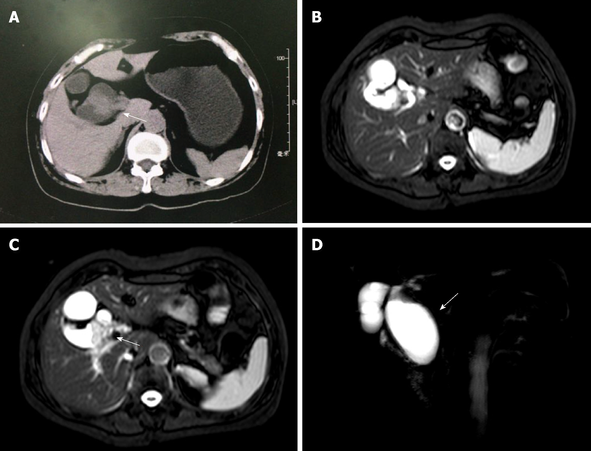
Figure 1 Computed tomography and magnetic resonance cholangiopancreatography findings.
A: Contrast-enhanced computed tomography showed a mass-like structure in the bile duct, and the malignant potential of the lesion could not be excluded (arrow); B: Magnetic resonance cholangiopancreatography (MRCP) revealed the dilated common bile duct; C: MRCP showed the space-occupying lesion in the hilar bile duct and the presence of a Klatskin tumor (arrow); D: Three-dimensional MRCP demonstrated a type I choledochal cyst (arrow).
- Citation: Lu BJ, Cao XD, Yuan N, Liu NN, Azami NL, Sun MY. Concomitant adenosquamous carcinoma and cystadenocarcinoma of the extrahepatic bile duct: A case report. World J Clin Cases 2019; 7(2): 215-220
- URL: https://www.wjgnet.com/2307-8960/full/v7/i2/215.htm
- DOI: https://dx.doi.org/10.12998/wjcc.v7.i2.215


