Copyright
©The Author(s) 2019.
World J Clin Cases. Jan 26, 2019; 7(2): 209-214
Published online Jan 26, 2019. doi: 10.12998/wjcc.v7.i2.209
Published online Jan 26, 2019. doi: 10.12998/wjcc.v7.i2.209
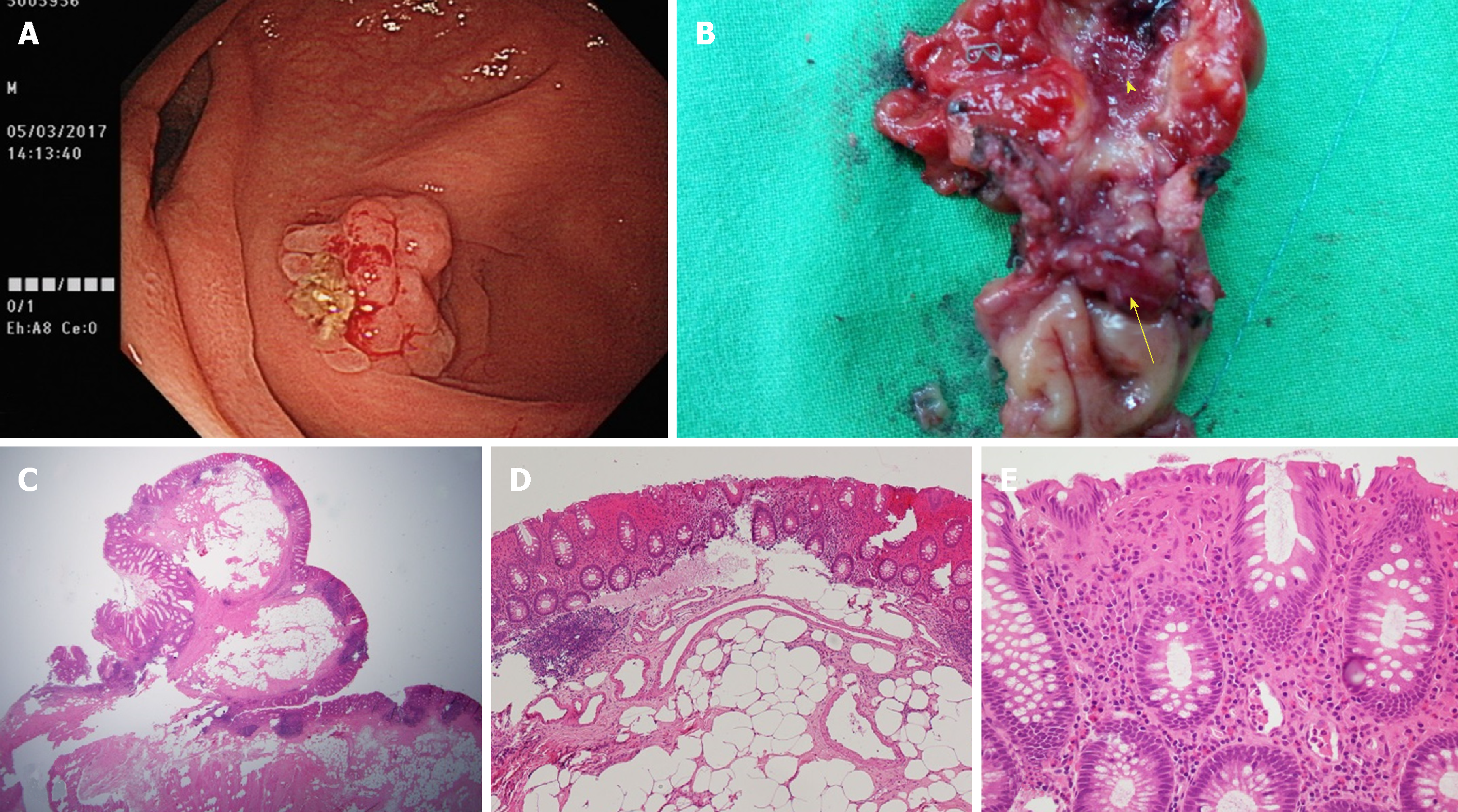
Figure 1 Images for the patient.
A: Colonoscopy revealing a polypoid mass occupying the appendiceal lumen; B: The opened specimen showing a lipoma located over the appendiceal orifice (arrow). Note the swollen and erythematous changes of the mucosa of the appendiceal lumen (arrowhead); C: Hematoxylin and eosin (HE) staining showing mature adipose cells inside the polypoid lesion (× 100); D: Mixed neutrophilic and lymphocytic infiltration noted in the mucosa covering the lesion (HE staining, × 200); E: Lymphocytic infiltration in the mucosa and lymphoid hyperplasia in the submucosa layer of the vermiform appendix (HE staining, × 400).
- Citation: Tsai KJ, Tai YS, Hung CM, Su YC. Cecal lipoma with subclinical appendicitis: A case report. World J Clin Cases 2019; 7(2): 209-214
- URL: https://www.wjgnet.com/2307-8960/full/v7/i2/209.htm
- DOI: https://dx.doi.org/10.12998/wjcc.v7.i2.209


