Copyright
©The Author(s) 2019.
World J Clin Cases. Jan 26, 2019; 7(2): 191-202
Published online Jan 26, 2019. doi: 10.12998/wjcc.v7.i2.191
Published online Jan 26, 2019. doi: 10.12998/wjcc.v7.i2.191
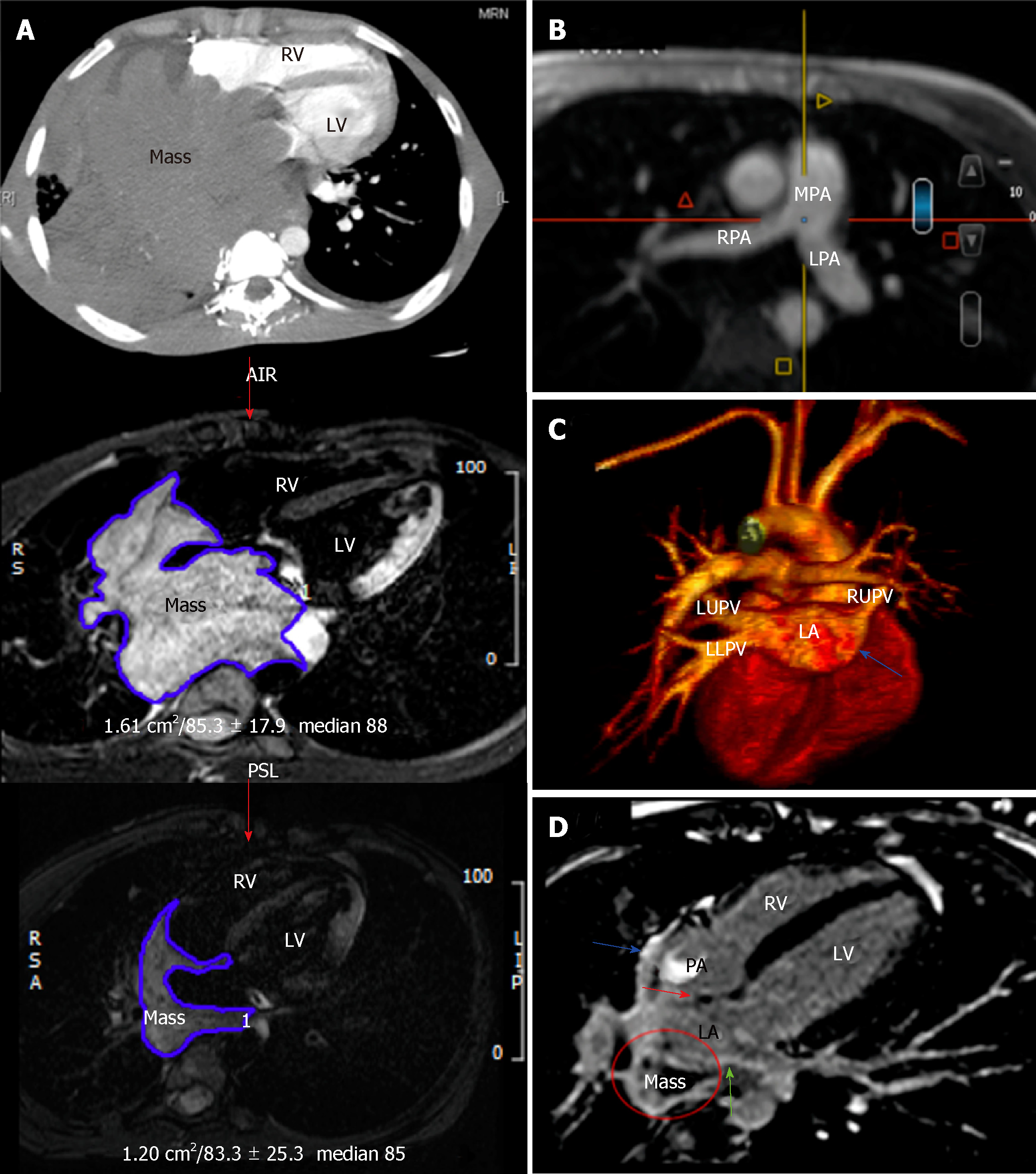
Figure 2 The patient’s imaging findings during the course of chemotherapy.
A: Follow-up computed tomography and CMRI at the heart level at consecutive time intervals showing gradual shrinkage of the mediastinal/cardiac mass size by 80% compared to baseline; B and C: Magnetic resonance angiography of the major cardiac vessels; B: Multiple-plane reformatted image showing the patency of the pulmonary arteries; C: In a 3-dimensional image, all pulmonary veins are now patent except the right lower pulmonary vein (blue arrow); D: Second CMRI-T1-weighted LGE image in a 4-chamber view revealing heterogeneous LGE of the mass (circle), suggesting tissue necrosis and fibrosis. The hyperintense signal of the interatrial septum (red arrow), left atrial (green arrow), and right atrial walls (blue arrows) suggest lymphoma resolution and healing by fibrosis. CMRI: Cardiac magnetic resonance imaging; RV: Right ventricle; LV: Left ventricle; RA: Right atrium; LA: Left atrium; LGE: Late gadolinium enhancement; MPA: Main pulmonary artery; RPA: Right pulmonary artery; LPA: Left pulmonary artery; LUPV: Left upper pulmonary vein; LLPV: Left lower pulmonary vein; RUPV: Right upper pulmonary vein.
- Citation: Al-Mehisen R, Al-Mohaissen M, Yousef H. Cardiac involvement in disseminated diffuse large B-cell lymphoma, successful management with chemotherapy dose reduction guided by cardiac imaging: A case report and review of literature. World J Clin Cases 2019; 7(2): 191-202
- URL: https://www.wjgnet.com/2307-8960/full/v7/i2/191.htm
- DOI: https://dx.doi.org/10.12998/wjcc.v7.i2.191


