Copyright
©The Author(s) 2019.
World J Clin Cases. Oct 6, 2019; 7(19): 3160-3167
Published online Oct 6, 2019. doi: 10.12998/wjcc.v7.i19.3160
Published online Oct 6, 2019. doi: 10.12998/wjcc.v7.i19.3160
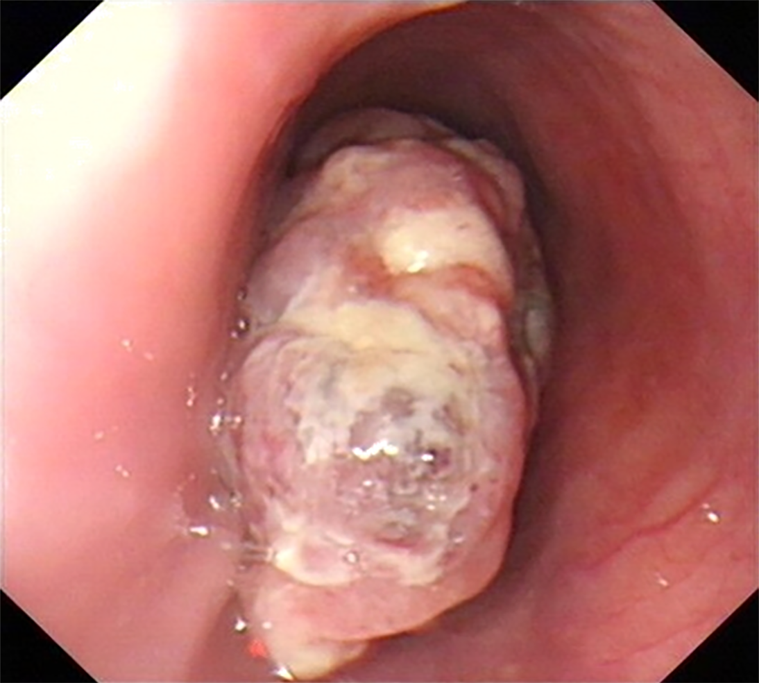
Figure 2 Upper gastrointestinal endoscopy.
A nonpigmented polypoid mass protruded into the esophageal lumen, located 30-35 cm from the incisors. The mass extended along the esophageal longitudinal axis.
- Citation: Zhang RX, Li YY, Liu CJ, Wang WN, Cao Y, Bai YH, Zhang TJ. Advanced primary amelanotic malignant melanoma of the esophagus: A case report. World J Clin Cases 2019; 7(19): 3160-3167
- URL: https://www.wjgnet.com/2307-8960/full/v7/i19/3160.htm
- DOI: https://dx.doi.org/10.12998/wjcc.v7.i19.3160


