Copyright
©The Author(s) 2019.
World J Clin Cases. Oct 6, 2019; 7(19): 3138-3144
Published online Oct 6, 2019. doi: 10.12998/wjcc.v7.i19.3138
Published online Oct 6, 2019. doi: 10.12998/wjcc.v7.i19.3138
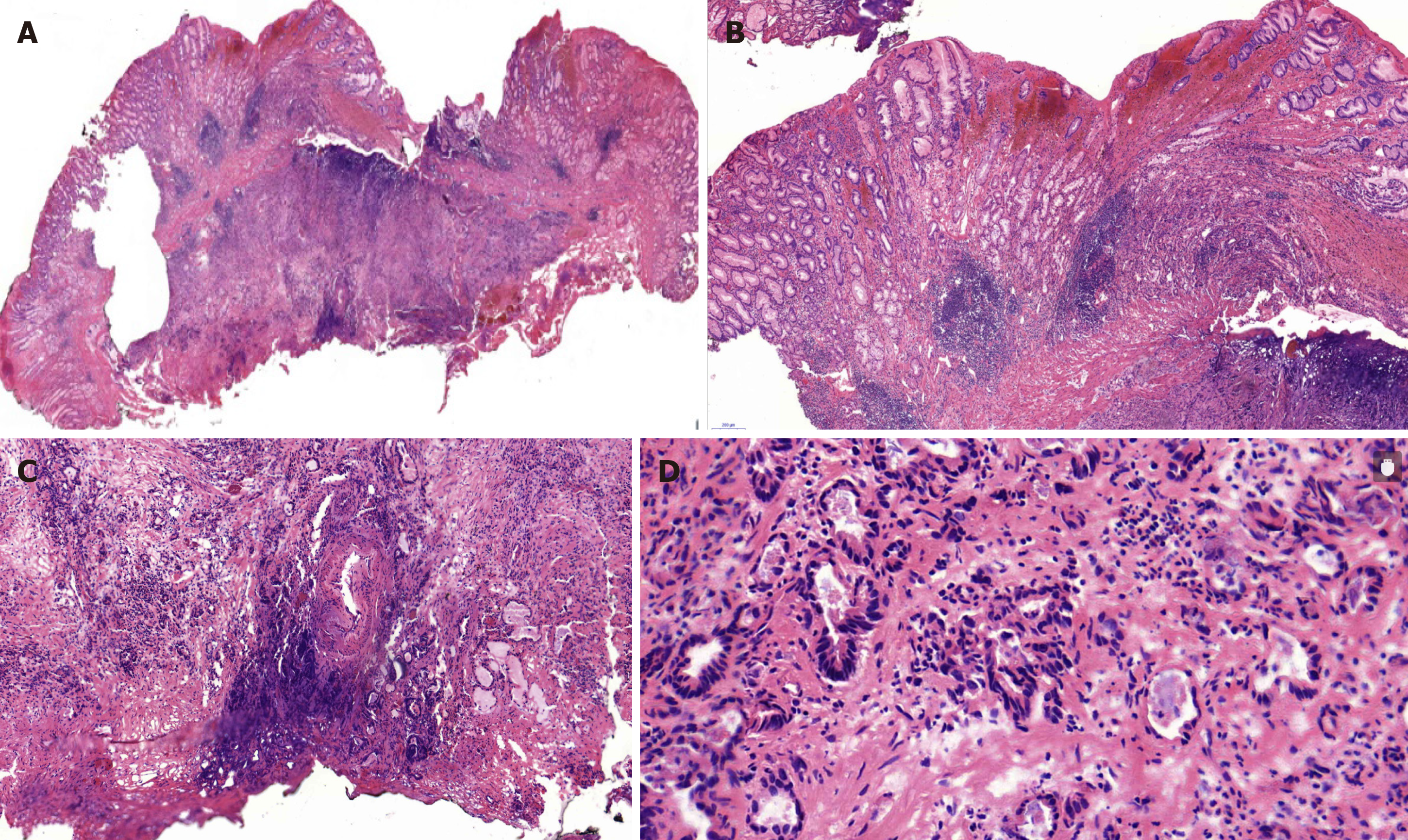
Figure 3 Histopathological findings (hematoxylin and eosin staining).
A moderately and poorly differentiated adenocarcinoma was suggested. A: The submucosal layer and muscularis mucosae were invaded; B: The tumor seemed to originate from the basal layer and invade towards the submucosal layer; C: The basal cutting edge was involved; D: Glands were arranged irregularly, with hyperchromatic nuclei and pathologic mitoses visible. Magnification: A: 40×; B and C: 100×; D: 200×.
- Citation: Cheng XL, Liu H. Gastric adenocarcinoma mimicking a submucosal tumor: A case report. World J Clin Cases 2019; 7(19): 3138-3144
- URL: https://www.wjgnet.com/2307-8960/full/v7/i19/3138.htm
- DOI: https://dx.doi.org/10.12998/wjcc.v7.i19.3138


