Copyright
©The Author(s) 2019.
World J Clin Cases. Oct 6, 2019; 7(19): 3090-3097
Published online Oct 6, 2019. doi: 10.12998/wjcc.v7.i19.3090
Published online Oct 6, 2019. doi: 10.12998/wjcc.v7.i19.3090
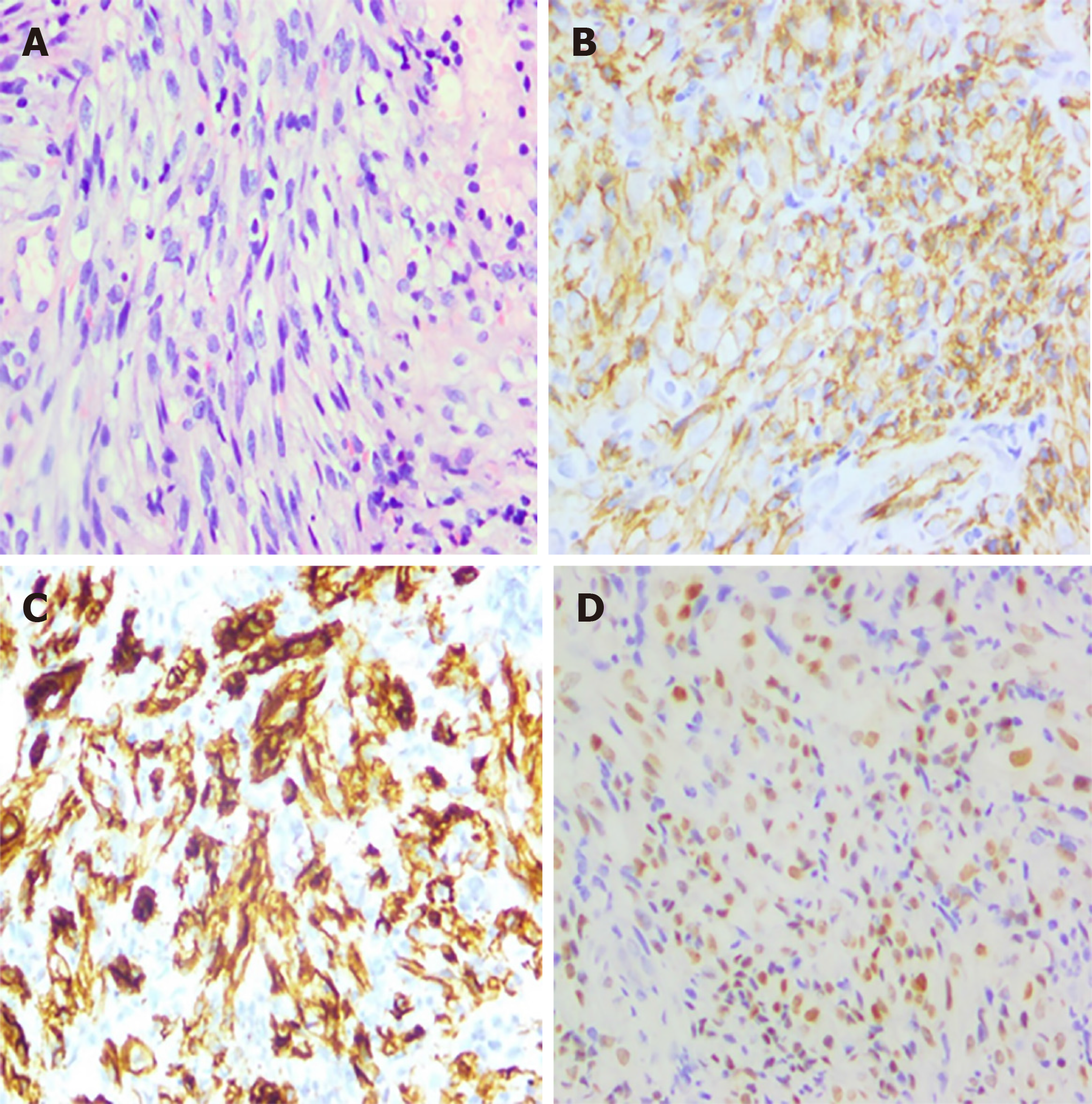
Figure 2 Haematoxylin and eosin stain and immunohistochemistry.
A: Proliferating spindle cells with severe atypia and a certain amount of inflammatory cells scattering among spindle cells (× 200); B: Immunohistochemical stains: positive for CD31, revealing the rectum with KS (× 200); C: Immunohistochemical stains: positive for CD34, revealing the rectum with KS (× 200); D: Immunohistochemical stains: positive for HHV8 antigen (× 200).
- Citation: Zhou QH, Guo YZ, Dai XH, Zhu B. Kaposi’s sarcoma manifested as lower gastrointestinal bleeding in a HIV/HBV-co-infected liver cirrhosis patient: A case report. World J Clin Cases 2019; 7(19): 3090-3097
- URL: https://www.wjgnet.com/2307-8960/full/v7/i19/3090.htm
- DOI: https://dx.doi.org/10.12998/wjcc.v7.i19.3090


