Copyright
©The Author(s) 2019.
World J Clin Cases. Oct 6, 2019; 7(19): 3033-3038
Published online Oct 6, 2019. doi: 10.12998/wjcc.v7.i19.3033
Published online Oct 6, 2019. doi: 10.12998/wjcc.v7.i19.3033
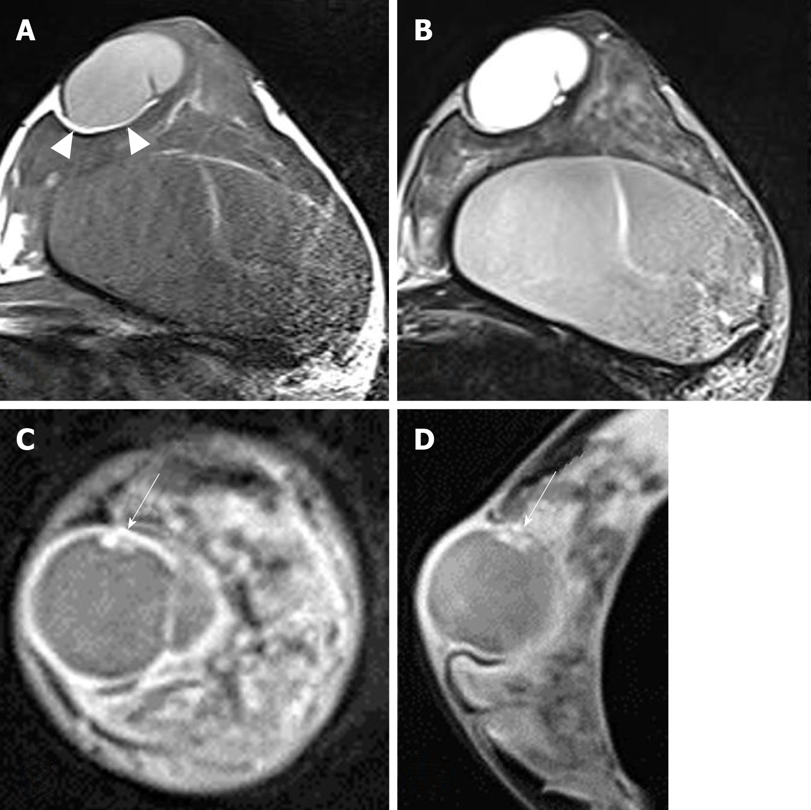
Figure 2 Breast magnetic resonance imaging showing a well-circumscribed oval mass of the left subareola.
A: The lesion attached to cutaneous layer of subareola and compressed the breast parenchyma. It showed a T1 hyper-intensity compared to signal in muscle. Thin fatty layer was seen between the mass and breast parenchyma (arrowheads). Breast implant was noted in the retromammary area; B: On T2-weighted axial image, the lesion showed a high signal intensity; C, D: On post-contrast fat saturation T1-weighted coronal (C) and sagittal (D) images, the mass showed a well-circumscribed thin and even enhancing wall. There was a small enhancing mural component in the inner wall of the mass (arrows).
- Citation: An JK, Woo JJ, Hong YO. Malignant sweat gland tumor of breast arising in pre-existing benign tumor: A case report. World J Clin Cases 2019; 7(19): 3033-3038
- URL: https://www.wjgnet.com/2307-8960/full/v7/i19/3033.htm
- DOI: https://dx.doi.org/10.12998/wjcc.v7.i19.3033


