Copyright
©The Author(s) 2019.
World J Clin Cases. Sep 26, 2019; 7(18): 2871-2878
Published online Sep 26, 2019. doi: 10.12998/wjcc.v7.i18.2871
Published online Sep 26, 2019. doi: 10.12998/wjcc.v7.i18.2871
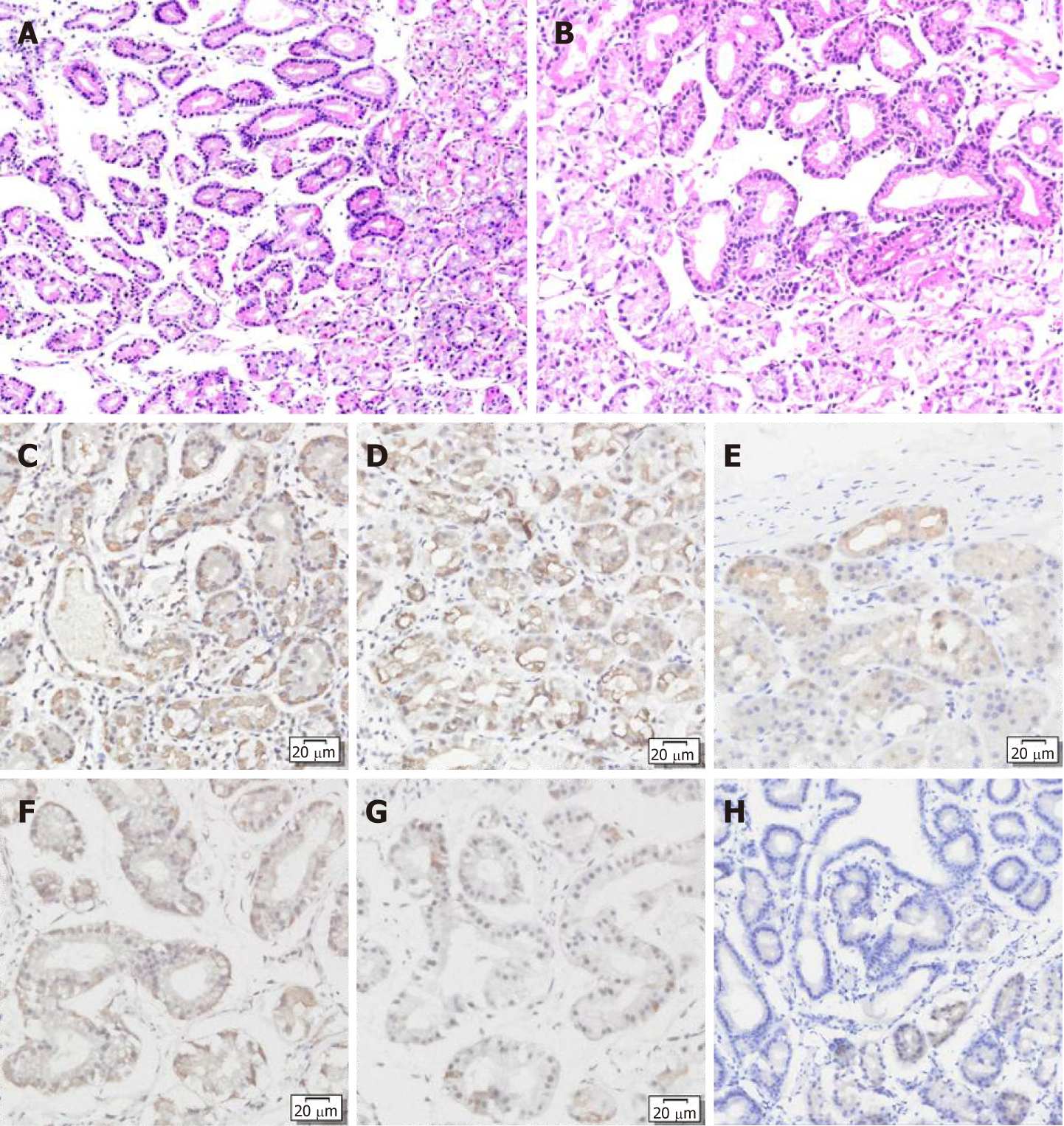
Figure 2 Hematoxylin and eosin staining showed that the tumors had clear demarcation from the surrounding fundus glands and an irregular glandular structure.
The tumors were composed of chief cell-like cells with mild nuclear atypia. A: Gastric corpus; B: Gastric fundus; C-H: Immunohistochemical staining showed that both lesions were positive for pepsinogen I (C and F) and MUC6 (D and G), and partially positive for H+/K+-ATPase (E and H).
- Citation: Chen O, Shao ZY, Qiu X, Zhang GP. Multiple gastric adenocarcinoma of fundic gland type: A case report. World J Clin Cases 2019; 7(18): 2871-2878
- URL: https://www.wjgnet.com/2307-8960/full/v7/i18/2871.htm
- DOI: https://dx.doi.org/10.12998/wjcc.v7.i18.2871


