Copyright
©The Author(s) 2019.
World J Clin Cases. Sep 26, 2019; 7(18): 2871-2878
Published online Sep 26, 2019. doi: 10.12998/wjcc.v7.i18.2871
Published online Sep 26, 2019. doi: 10.12998/wjcc.v7.i18.2871
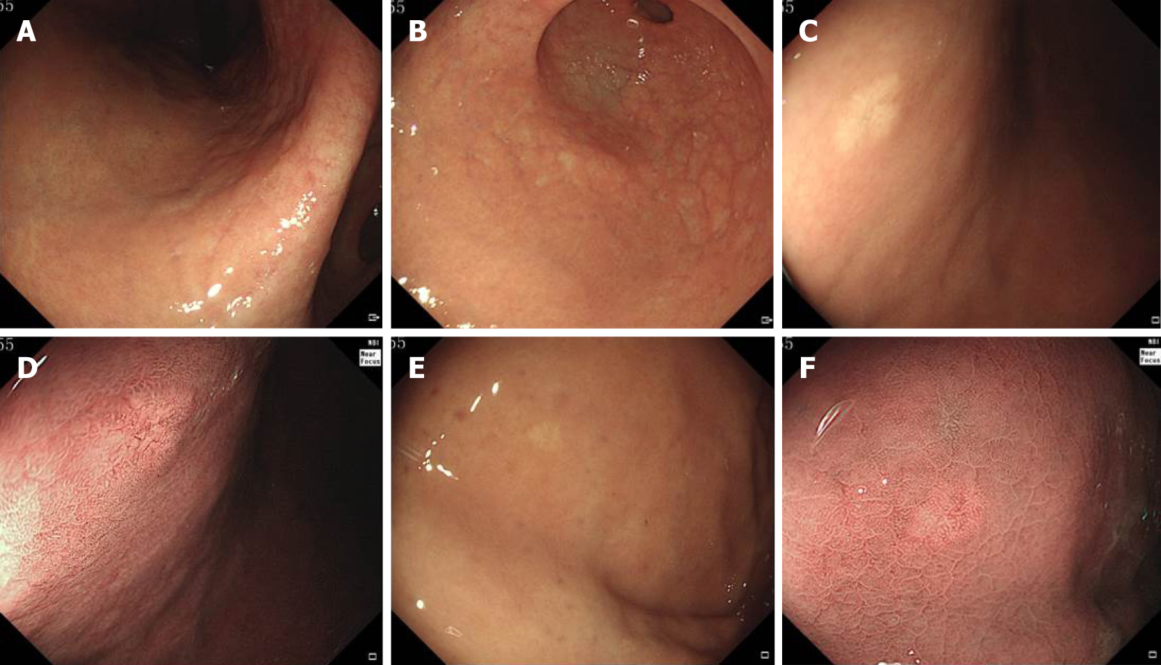
Figure 1 Endoscopic findings of all lesions.
A and B: The gastric mucosa showed grade C-2 atrophic gastritis according to the Kimura-Takemoto classification; C: White light endoscopy revealed a submucosal tumor-like elevated mass with a whitish mucosal surface on the anterior gastric corpus wall; D: Narrow-band imaging showed an irregular microvascular pattern and dilatation of microvessels with branching architecture; E: White light endoscopy revealed a flat shaped lesion with a whitish mucosal surface on the gastric fundus; F: Narrow-band imaging showed regular and dilated microvessels.
- Citation: Chen O, Shao ZY, Qiu X, Zhang GP. Multiple gastric adenocarcinoma of fundic gland type: A case report. World J Clin Cases 2019; 7(18): 2871-2878
- URL: https://www.wjgnet.com/2307-8960/full/v7/i18/2871.htm
- DOI: https://dx.doi.org/10.12998/wjcc.v7.i18.2871


