Copyright
©The Author(s) 2019.
World J Clin Casesol. Sep 26, 2019; 7(18): 2864-2870
Published online Sep 26, 2019. doi: 10.12998/wjcc.v7.i18.2864
Published online Sep 26, 2019. doi: 10.12998/wjcc.v7.i18.2864
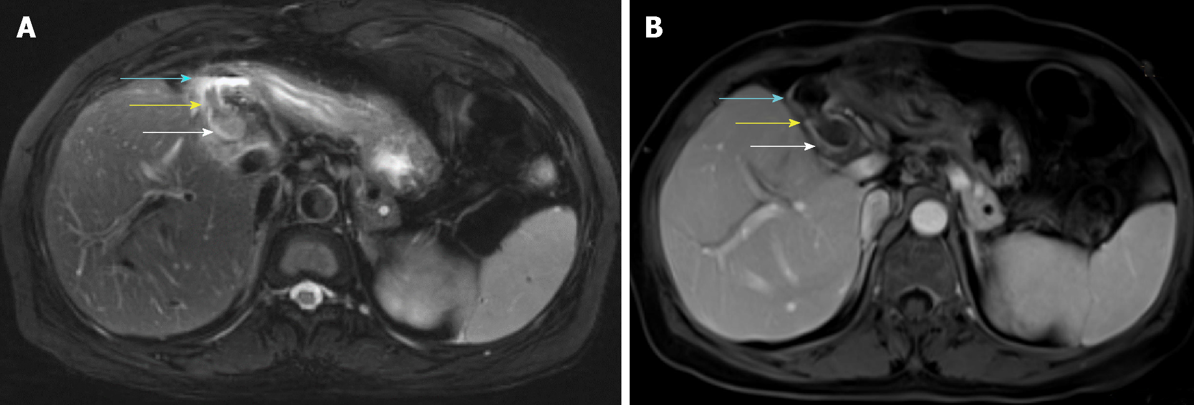
Figure 1 Magnetic resonance imaging.
A: Axial T2 weighted image showing a fistula connecting the bile duct (white arrow) and the duodenal bulb (blue arrow) which had a gas-liquid plane inside. Inside the fistula, there was a hypointense lesion (yellow arrow); B: Axial T1 weighted image contrast enhanced showing no enhancement of the bile duct (white arrow), duodenal bulb (blue arrow), or lesion (yellow arrow).
- Citation: Yang S, Yang L, Wang XY, Yang YM. Endoscopic mucosal resection of a bile duct polyp: A case report. World J Clin Casesol 2019; 7(18): 2864-2870
- URL: https://www.wjgnet.com/2307-8960/full/v7/i18/2864.htm
- DOI: https://dx.doi.org/10.12998/wjcc.v7.i18.2864


