Copyright
©The Author(s) 2019.
World J Clin Cases. Sep 26, 2019; 7(18): 2831-2837
Published online Sep 26, 2019. doi: 10.12998/wjcc.v7.i18.2831
Published online Sep 26, 2019. doi: 10.12998/wjcc.v7.i18.2831
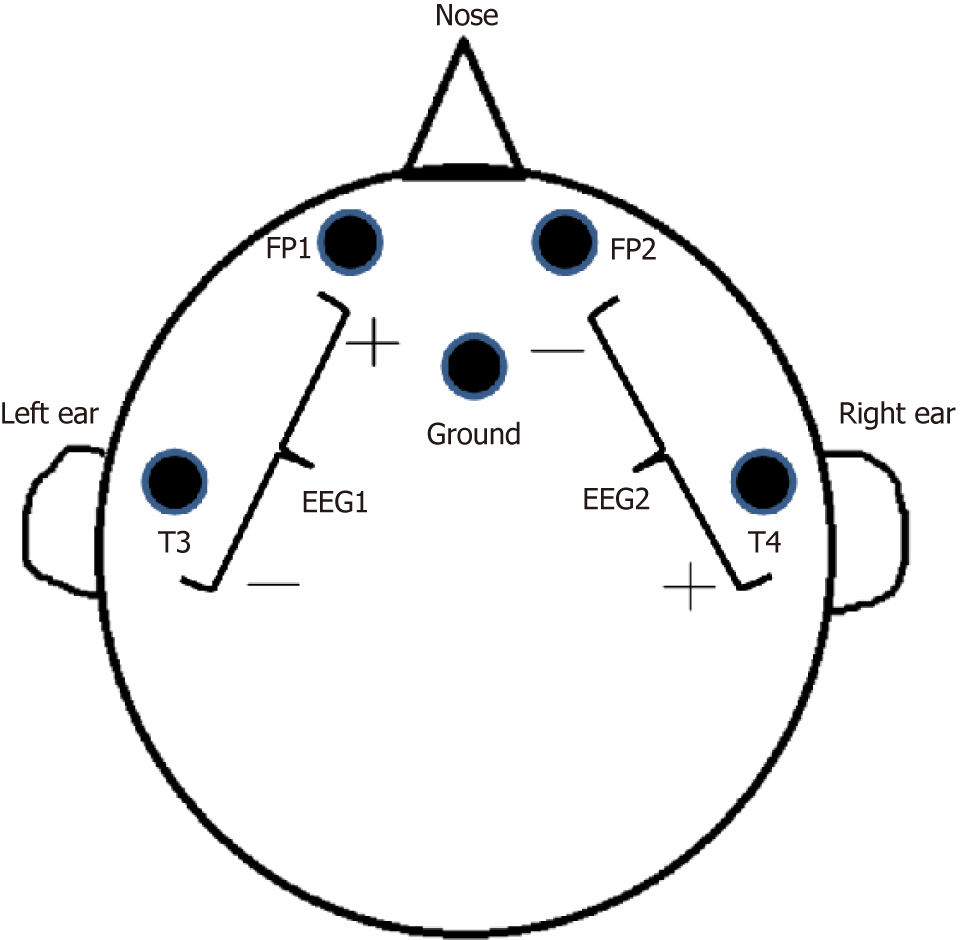
Figure 2 The positions of five electrodes on the scalp.
EEG: Electroencephalogram; FP1: Front point 1, 1 cm above the midline of left eyebrow; FP2: Front point 2, 1 cm above the midline of right eyebrow; T3: Temporal point 3, 1 cm above the left ear tip; T4: Temporal point 4, 1 cm above the right ear tip; Ground: Ground electrode, 1 cm above the nasion.
- Citation: Hor S, Chen CY, Tsai ST. Propofol pump controls nonconvulsive status epilepticus in a hepatic encephalopathy patient: A case report. World J Clin Cases 2019; 7(18): 2831-2837
- URL: https://www.wjgnet.com/2307-8960/full/v7/i18/2831.htm
- DOI: https://dx.doi.org/10.12998/wjcc.v7.i18.2831


