Copyright
©The Author(s) 2019.
World J Clin Cases. Sep 26, 2019; 7(18): 2823-2830
Published online Sep 26, 2019. doi: 10.12998/wjcc.v7.i18.2823
Published online Sep 26, 2019. doi: 10.12998/wjcc.v7.i18.2823
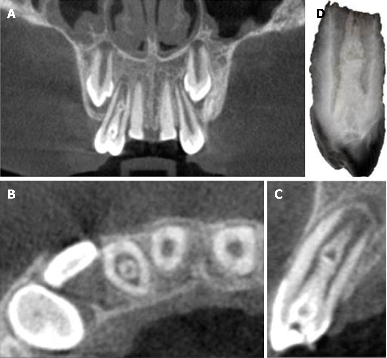
Figure 3 Cone-beam computed tomography images.
A: Coronal view shows a central bulge; B: Axial view shows the relative positions of the main root canal and pseudo root canal; C: Sagittal plane view indicates that the enlargement is connected with the main root canal; D: Reconstruction of the three-dimensional structure of the dens invaginatus.
- Citation: Lee HN, Chen YK, Chen CH, Huang CY, Su YH, Huang YW, Chuang FH. Conservative pulp treatment for Oehlers type III dens invaginatus: A case report. World J Clin Cases 2019; 7(18): 2823-2830
- URL: https://www.wjgnet.com/2307-8960/full/v7/i18/2823.htm
- DOI: https://dx.doi.org/10.12998/wjcc.v7.i18.2823


