Copyright
©The Author(s) 2019.
World J Clin Cases. Sep 26, 2019; 7(18): 2815-2822
Published online Sep 26, 2019. doi: 10.12998/wjcc.v7.i18.2815
Published online Sep 26, 2019. doi: 10.12998/wjcc.v7.i18.2815
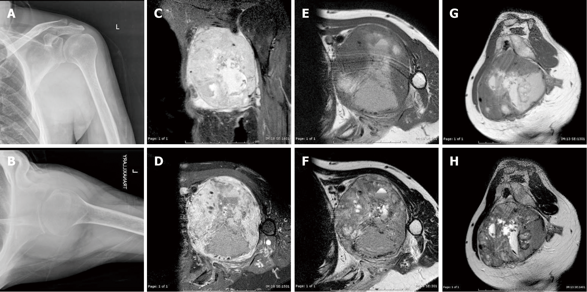
Figure 3 Radiographs and preoperative magnetic resonance imaging.
A, B: Preoperative plain coronal (A) and trans-scapular (B) radiographs of the left shoulder showing a large soft tissue shadow in the axilla; C-H: Preoperative magnetic resonance imaging: coronal (C) and transverse (D) fat suppressed T1-weighted images after intravenous administration of gadolinium, transverse T1-weighted (E), transverse T2-weighted (F), sagittal T1-weighted (G), and sagittal T2-weighted (H) images demonstrating a large heterogeneous, contrast-enhancing mass bulging from the posterior cord of the brachial plexus, encasing the axillary artery and vein, and occupying the entire axillary space.
- Citation: Thanindratarn P, Chobpenthai T, Phorkhar T, Nelson SD. Glomus tumor of uncertain malignant potential of the brachial plexus: A case report. World J Clin Cases 2019; 7(18): 2815-2822
- URL: https://www.wjgnet.com/2307-8960/full/v7/i18/2815.htm
- DOI: https://dx.doi.org/10.12998/wjcc.v7.i18.2815


