Copyright
©The Author(s) 2019.
World J Clin Cases. Sep 26, 2019; 7(18): 2794-2801
Published online Sep 26, 2019. doi: 10.12998/wjcc.v7.i18.2794
Published online Sep 26, 2019. doi: 10.12998/wjcc.v7.i18.2794
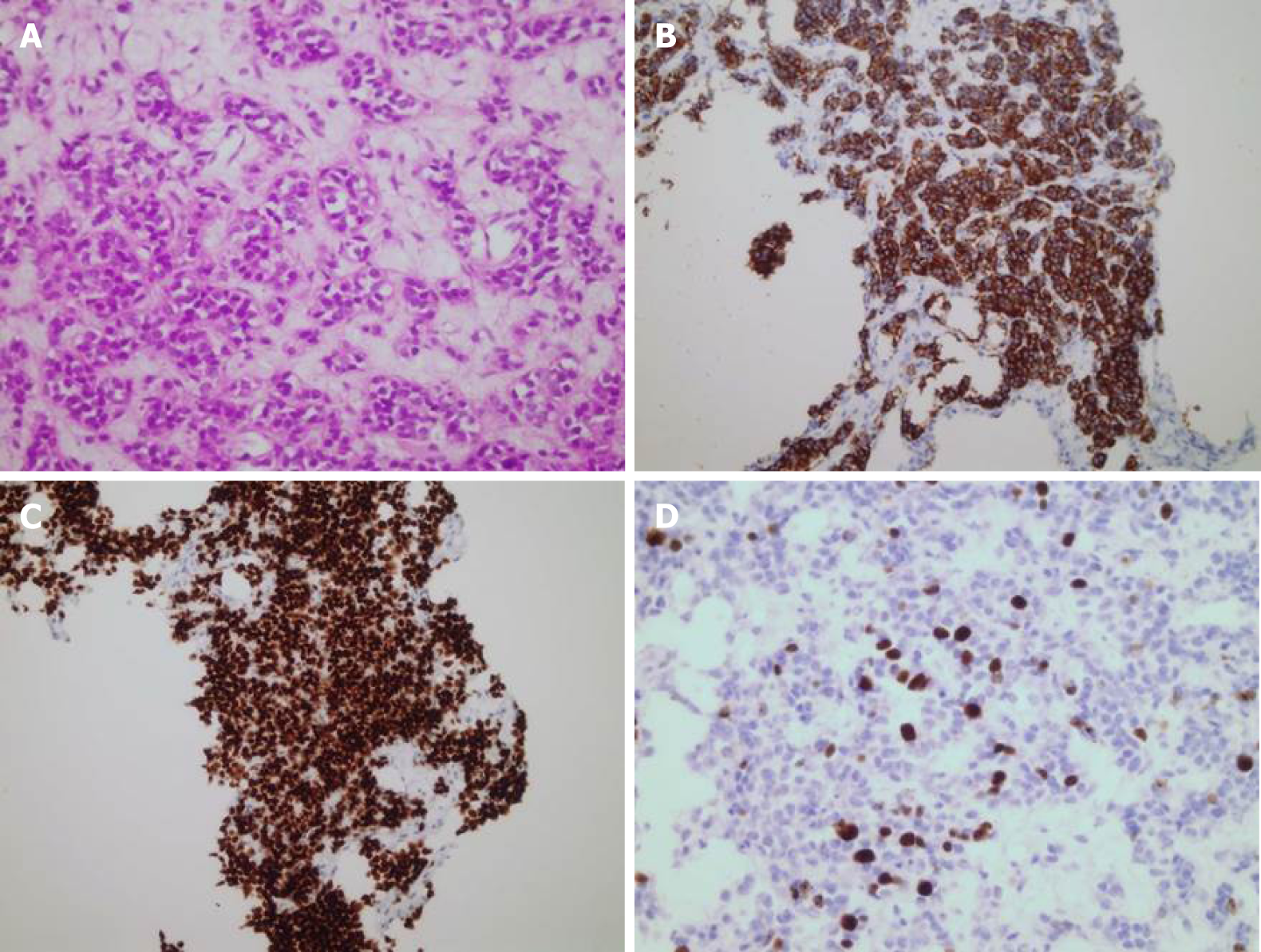
Figure 3 Liver biopsy and immunohistochemistry of neuroendocrine tumor.
A: Closely packed nests of well-differentiated neuroendocrine cells (hematoxylin and eosin staining; magnification 400 ×); B: Synaptophysin staining (magnification 200 ×); C: TTF-1 staining (magnification 200 ×); D: Ki67 (magnification 400 ×).
- Citation: Mrzljak A, Kocman B, Skrtic A, Furac I, Popic J, Franusic L, Zunec R, Mayer D, Mikulic D. Liver re-transplantation for donor-derived neuroendocrine tumor: A case report. World J Clin Cases 2019; 7(18): 2794-2801
- URL: https://www.wjgnet.com/2307-8960/full/v7/i18/2794.htm
- DOI: https://dx.doi.org/10.12998/wjcc.v7.i18.2794


