Copyright
©The Author(s) 2019.
World J Clin Cases. Sep 26, 2019; 7(18): 2794-2801
Published online Sep 26, 2019. doi: 10.12998/wjcc.v7.i18.2794
Published online Sep 26, 2019. doi: 10.12998/wjcc.v7.i18.2794
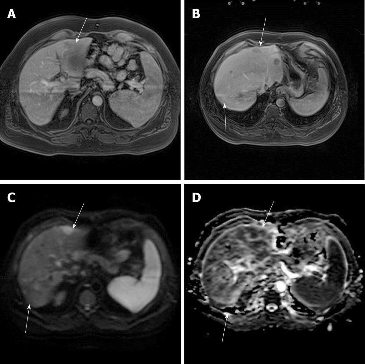
Figure 1 MR images 11 mo after LT.
A: Axial postcontrast T1 MR images showed light peripheral enhancement of the lesion in segment III in the arterial phase (arrow); B: Postcontrast axial images through the transplanted liver revealed hypointensive lesions in segments III and VIII in the portal venous phase (arrows); C: Diffusion-weighted images with b 600 showed hyperintensity of the same lesions; D: On apparent diffusion coefficient map, the lesions showed diffusion restriction. LT: Liver transplant; MR: Magnetic resonance.
- Citation: Mrzljak A, Kocman B, Skrtic A, Furac I, Popic J, Franusic L, Zunec R, Mayer D, Mikulic D. Liver re-transplantation for donor-derived neuroendocrine tumor: A case report. World J Clin Cases 2019; 7(18): 2794-2801
- URL: https://www.wjgnet.com/2307-8960/full/v7/i18/2794.htm
- DOI: https://dx.doi.org/10.12998/wjcc.v7.i18.2794


