Copyright
©The Author(s) 2019.
World J Clin Cases. Sep 6, 2019; 7(17): 2637-2643
Published online Sep 6, 2019. doi: 10.12998/wjcc.v7.i17.2637
Published online Sep 6, 2019. doi: 10.12998/wjcc.v7.i17.2637
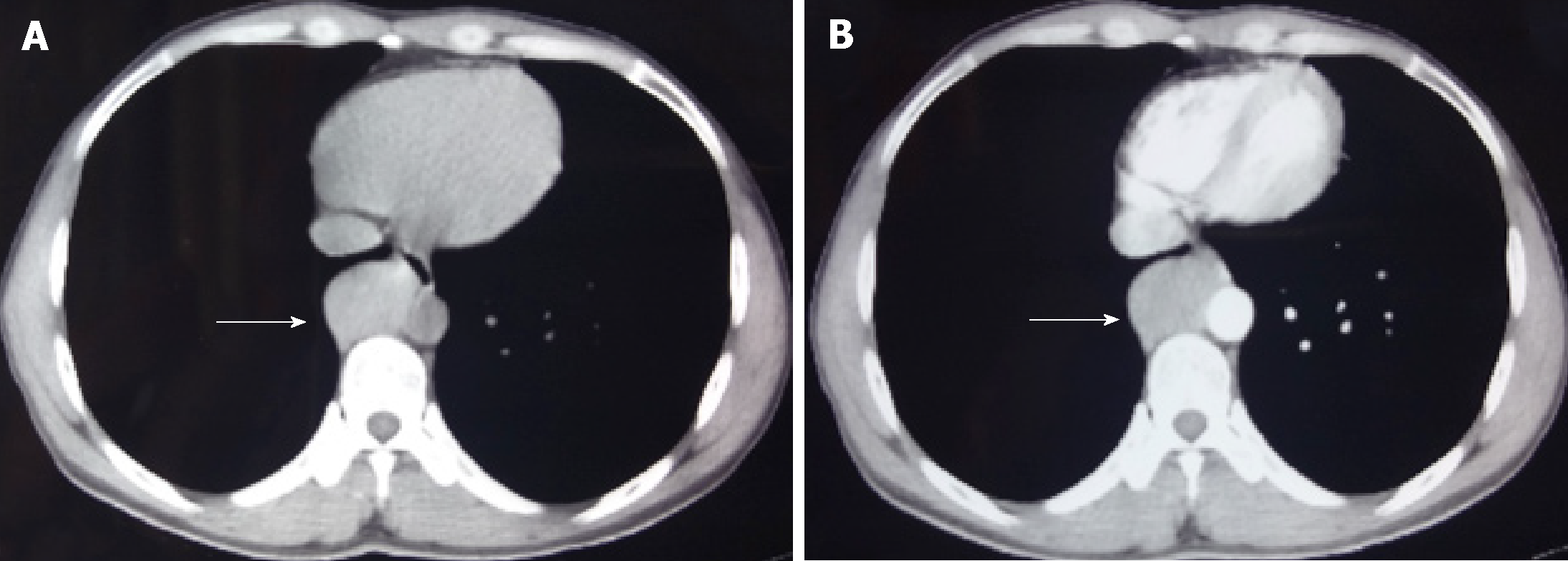
Figure 1 Chest computed tomography.
The oval tumor (shown by the arrows) was located on the lateral wall of the abdominal aortic artery, with uniform density and a size of about 4.5 cm × 3.0 cm. The boundary with surrounding tissues is clear. The computed tomography (CT) value of the flat scan is about 57 HU (A), and the enhanced CT value is about 113 HU (B).
- Citation: Qi DJ, Zhang QF. Calcifying fibrous tumor of the mediastinum: A case report. World J Clin Cases 2019; 7(17): 2637-2643
- URL: https://www.wjgnet.com/2307-8960/full/v7/i17/2637.htm
- DOI: https://dx.doi.org/10.12998/wjcc.v7.i17.2637


