Copyright
©The Author(s) 2019.
World J Clin Cases. Sep 6, 2019; 7(17): 2623-2629
Published online Sep 6, 2019. doi: 10.12998/wjcc.v7.i17.2623
Published online Sep 6, 2019. doi: 10.12998/wjcc.v7.i17.2623
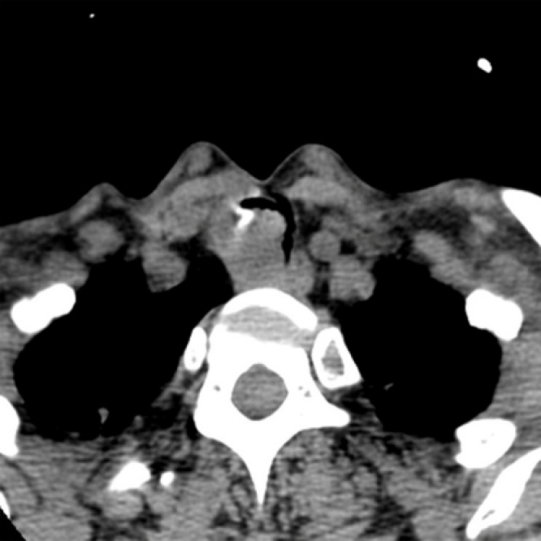
Figure 1 Computed tomography scan showing an inhomogeneous, broad-based lesion arising from the tracheal wall on the right side.
Calcification can be seen in the anterior part of the tumor.
- Citation: Gao HX, Li Q, Chang WL, Zhang YL, Wang XZ, Zou XX. Carcinoma ex pleomorphic adenoma of the trachea: A case report. World J Clin Cases 2019; 7(17): 2623-2629
- URL: https://www.wjgnet.com/2307-8960/full/v7/i17/2623.htm
- DOI: https://dx.doi.org/10.12998/wjcc.v7.i17.2623


