Copyright
©The Author(s) 2019.
World J Clin Cases. Sep 6, 2019; 7(17): 2605-2610
Published online Sep 6, 2019. doi: 10.12998/wjcc.v7.i17.2605
Published online Sep 6, 2019. doi: 10.12998/wjcc.v7.i17.2605
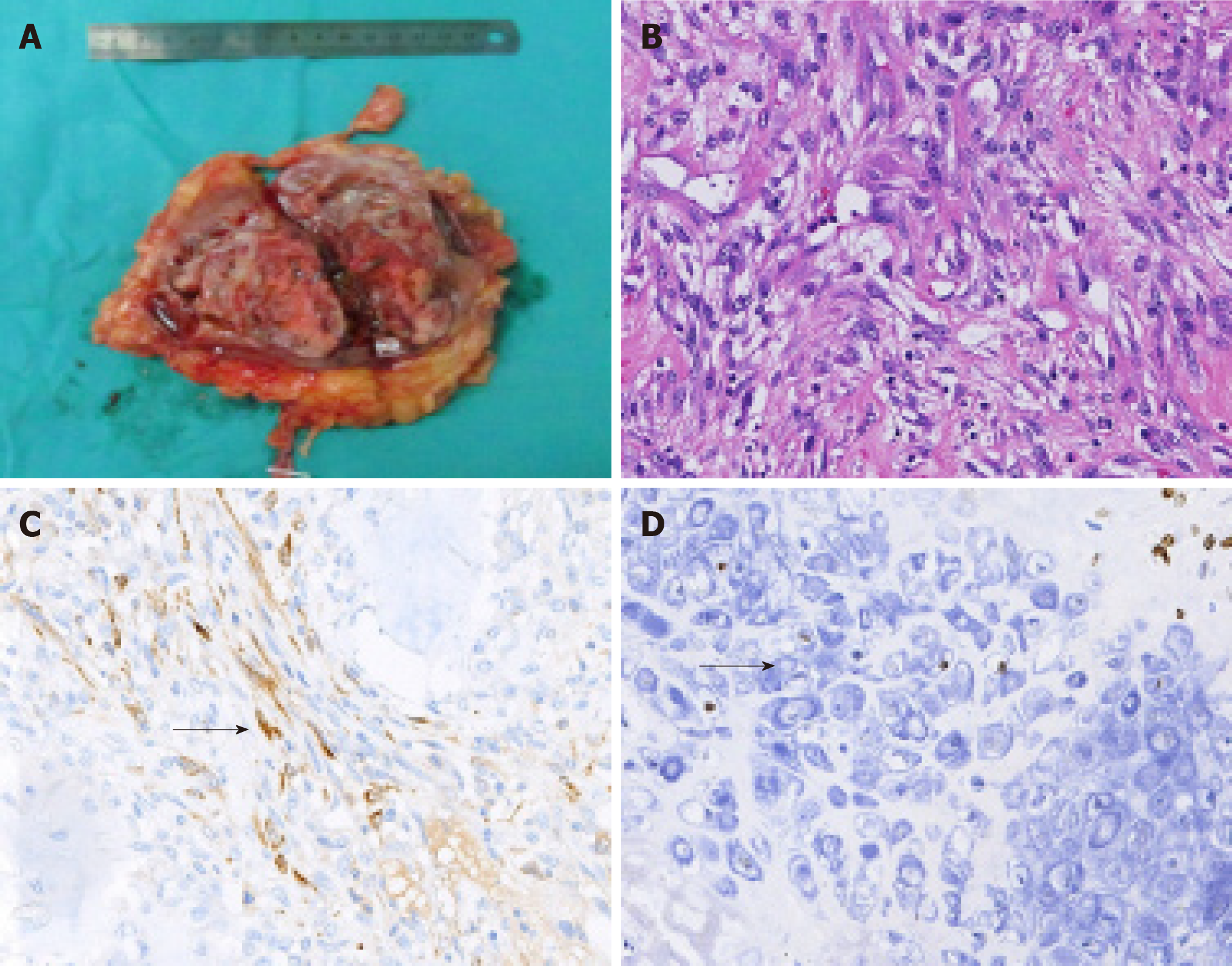
Figure 2 Gross specimen and histological examination of our case of renal organ-associated pseudosarcomatous myofibroblastic proliferation with ossification.
A: Longitudinal section of our patient’s kidney showing a gray-white and pink-grey mass, with capsule in the lower pole of the kidney; B: Microscopic view of the renal organ-associated pseudosarcomatous myofibroblastic proliferation staining with hematoxylin eosin (original magnification × 200). The lesions were found to be composed mainly of proliferative fusiform myofibroblasts and inflammatory cells; C, D: Immunohistochemical staining showed positivity for calponin (C) and SATB-2 (D) (Arrows indicate positive cells).
- Citation: Zhai TY, Luo BJ, Jia ZK, Zhang ZG, Li X, Li H, Yang JJ. Organ-associated pseudosarcomatous myofibroblastic proliferation with ossification in the lower pole of the kidney mimicking renal pelvic carcinoma: A case report. World J Clin Cases 2019; 7(17): 2605-2610
- URL: https://www.wjgnet.com/2307-8960/full/v7/i17/2605.htm
- DOI: https://dx.doi.org/10.12998/wjcc.v7.i17.2605


