Copyright
©The Author(s) 2019.
World J Clin Cases. Sep 6, 2019; 7(17): 2605-2610
Published online Sep 6, 2019. doi: 10.12998/wjcc.v7.i17.2605
Published online Sep 6, 2019. doi: 10.12998/wjcc.v7.i17.2605
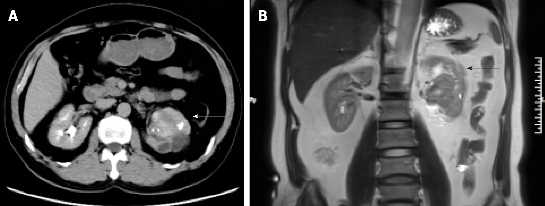
Figure 1 Imaging examination of our case of renal organ-associated pseudosarcomatous myofibroblastic proliferation.
A: Computed tomography showed a heterogeneous mass (indicated with arrows) of 6 cm in diameter at the left pyeloureteral junction; B: Magnetic resonance imaging showed that the left renal pelvis was occupied by a cystic and solid mass (indicated with arrows) with calcification or ossification.
- Citation: Zhai TY, Luo BJ, Jia ZK, Zhang ZG, Li X, Li H, Yang JJ. Organ-associated pseudosarcomatous myofibroblastic proliferation with ossification in the lower pole of the kidney mimicking renal pelvic carcinoma: A case report. World J Clin Cases 2019; 7(17): 2605-2610
- URL: https://www.wjgnet.com/2307-8960/full/v7/i17/2605.htm
- DOI: https://dx.doi.org/10.12998/wjcc.v7.i17.2605


