Copyright
©The Author(s) 2019.
World J Clin Cases. Sep 6, 2019; 7(17): 2587-2596
Published online Sep 6, 2019. doi: 10.12998/wjcc.v7.i17.2587
Published online Sep 6, 2019. doi: 10.12998/wjcc.v7.i17.2587
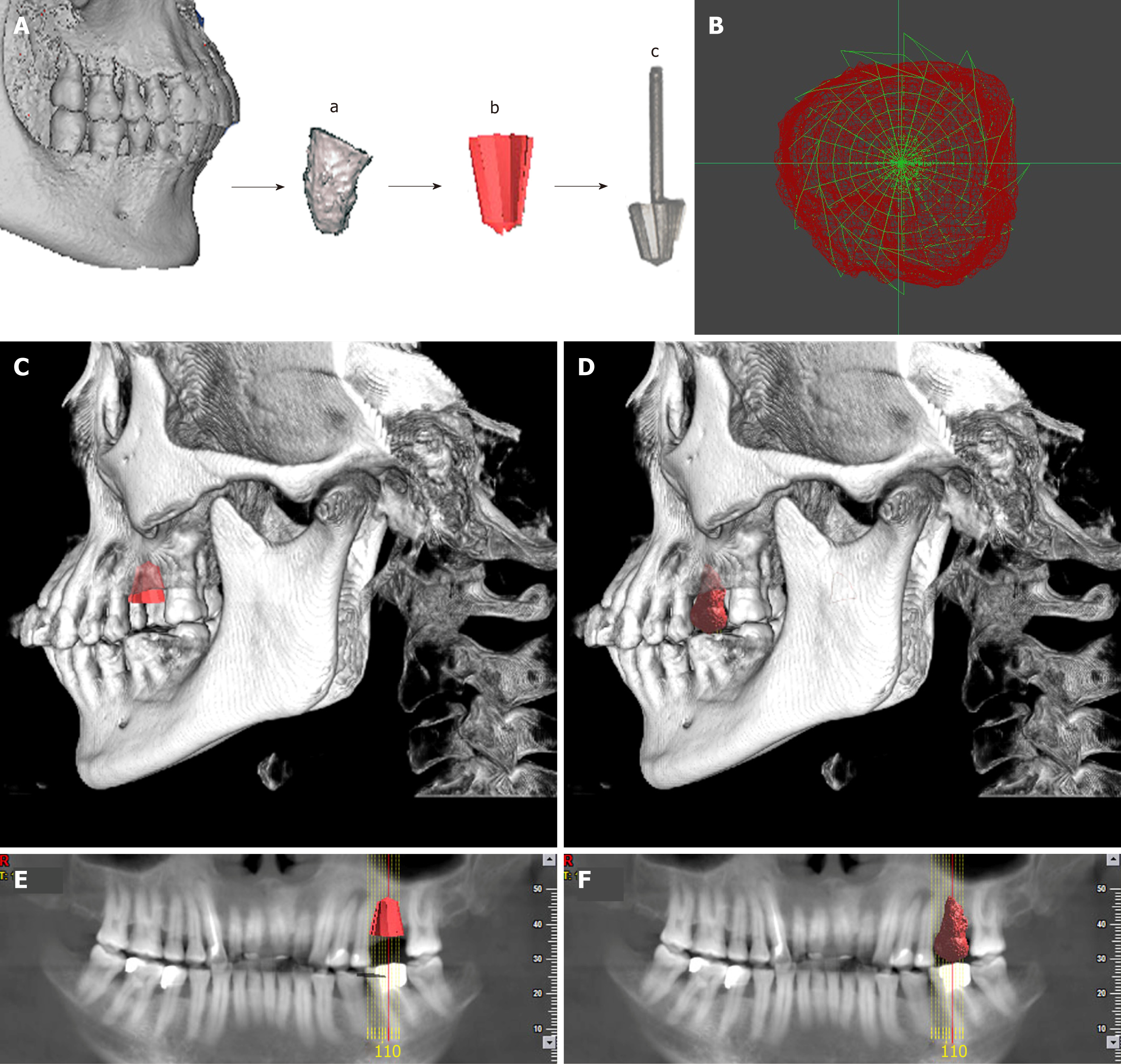
Figure 2 Individual drill compared with donor’s tooth.
A: The 3D image of the donor’s tooth was extracted from the cone beam computed tomography, a computer-aided design model was built using measuring instrument to gather information for the shape and size, and an individual drill was manufactured by direct metal laser sintering; B: The cross section comparison of the individual drill and the donor’s tooth; C and E: The model of the drill in the recipient’s alveolar bone; D and F: The model of the donor’s tooth in the recipient’s alveolar bone.
- Citation: Xu HD, Miron RJ, Zhang XX, Zhang YF. Allogenic tooth transplantation using 3D printing: A case report and review of the literature. World J Clin Cases 2019; 7(17): 2587-2596
- URL: https://www.wjgnet.com/2307-8960/full/v7/i17/2587.htm
- DOI: https://dx.doi.org/10.12998/wjcc.v7.i17.2587


