Copyright
©The Author(s) 2019.
World J Clin Cases. Sep 6, 2019; 7(17): 2580-2586
Published online Sep 6, 2019. doi: 10.12998/wjcc.v7.i17.2580
Published online Sep 6, 2019. doi: 10.12998/wjcc.v7.i17.2580
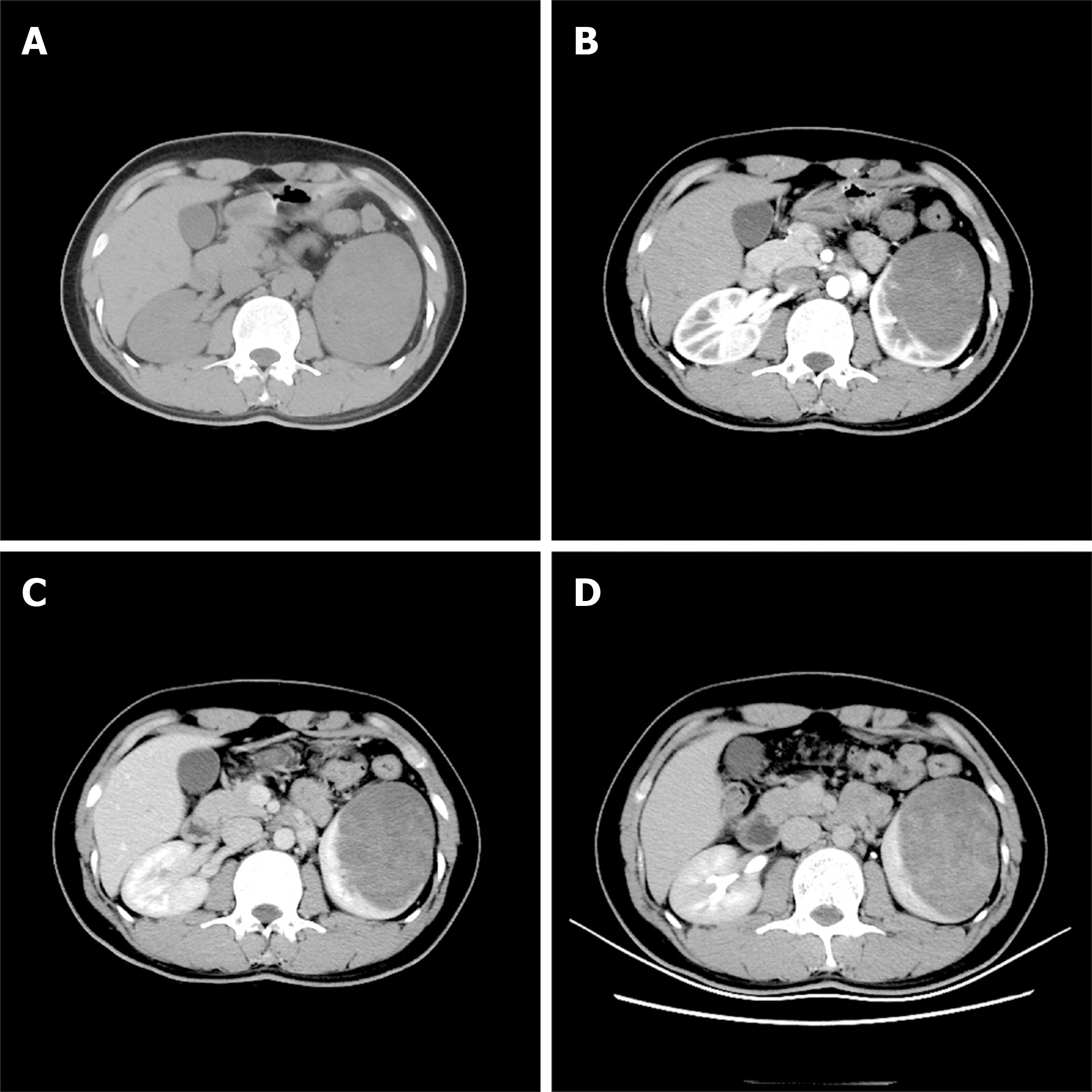
Figure 1 Computed tomography.
A: Non-enhanced computed tomography image showing a round, well-defined, and mixed mass; B: Enhanced computed tomography image during the corticomedullary phase; C: Nephrographic phase; D: Delayed enhancement of the solid part and no enhancement of the cystic part.
- Citation: Ye J, Xu Q, Zheng J, Wang SA, Wu YW, Cai JH, Yuan H. Imaging of mixed epithelial and stromal tumor of the kidney: A case report and review of the literature. World J Clin Cases 2019; 7(17): 2580-2586
- URL: https://www.wjgnet.com/2307-8960/full/v7/i17/2580.htm
- DOI: https://dx.doi.org/10.12998/wjcc.v7.i17.2580


