Copyright
©The Author(s) 2019.
World J Clin Cases. Sep 6, 2019; 7(17): 2526-2535
Published online Sep 6, 2019. doi: 10.12998/wjcc.v7.i17.2526
Published online Sep 6, 2019. doi: 10.12998/wjcc.v7.i17.2526
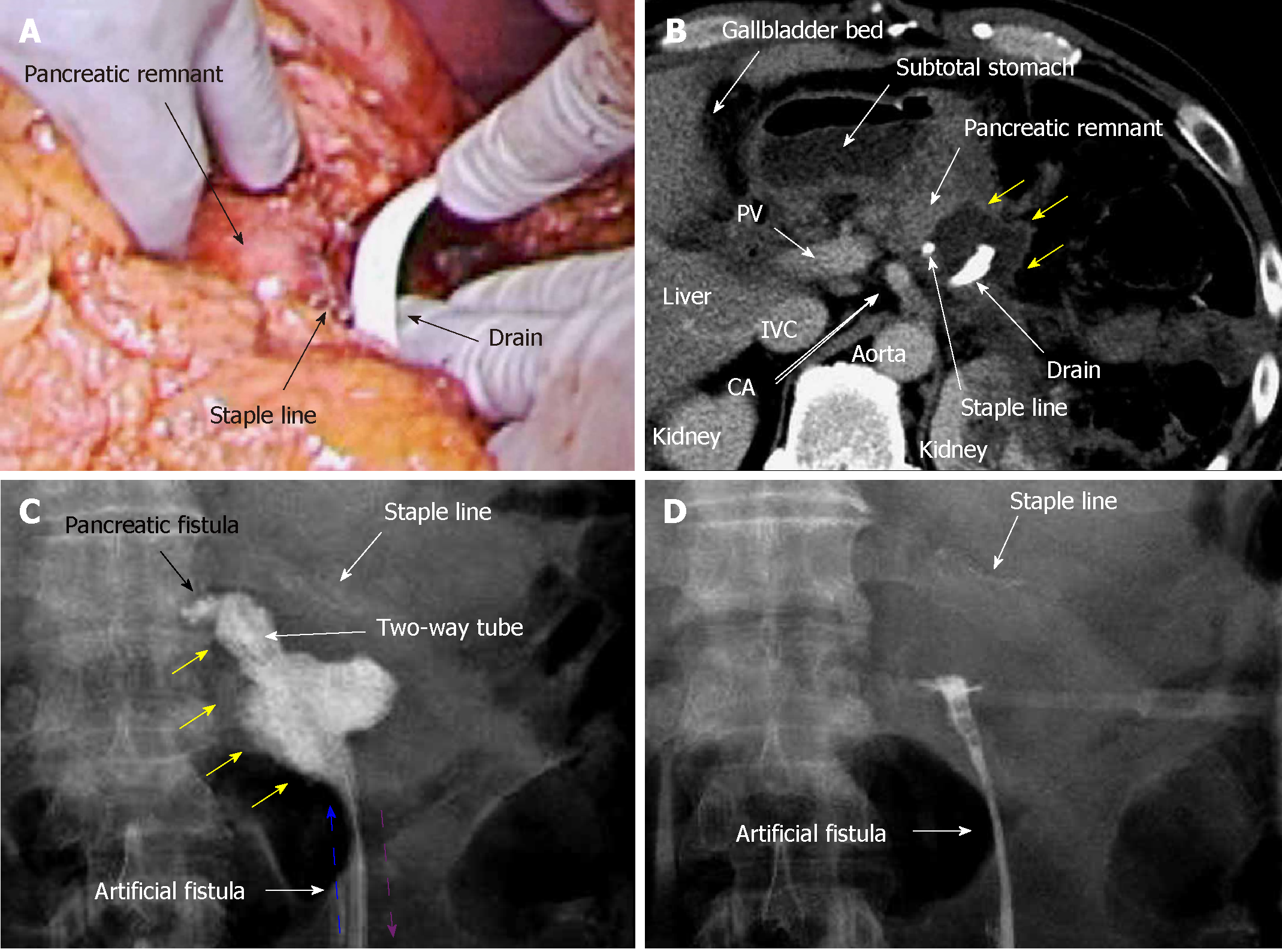
Figure 3 Case 2.
A: Pancreatectomy was performed using a linear stapler, and we placed a drain near the staple line; B: Contrast computed tomography on postoperative day (POD) 9 revealed that intractable pancreatic fistula had led to an intraperitoneal abscess near the staple line (yellow arrows); C: Fistulography via the drain on POD 9 revealed intractable pancreatic fistula (black arrow) and the abscess cavity (yellow arrows). From POD 9, saline irrigation (blue arrow) was continuously injected into the abscess cavity via a two-way tube with the drainage route (purple arrow). Thereafter, amylase levels in the drainage discharge decreased immediately; D: Fistulography on POD 12 revealed that the abscess resolved after 4 d of continuous local lavage. CHA: Common hepatic artery; CLL: Continuous local lavage; IVC: Inferior vena cava; POD: Postoperative day.
- Citation: Hori T, Ogawa K, Yamamoto H, Harada H, Matsumura K, Yamamoto M, Yamada M, Yazawa T, Kuriyama K, Tani M, Yasukawa D, Kamada Y, Aisu Y, Tani R, Aoyama R, Nakayama S, Sasaki Y, Nishimoto K, Zaima M. Impact of continuous local lavage on pancreatic juice-related postoperative complications: Three case reports. World J Clin Cases 2019; 7(17): 2526-2535
- URL: https://www.wjgnet.com/2307-8960/full/v7/i17/2526.htm
- DOI: https://dx.doi.org/10.12998/wjcc.v7.i17.2526


