Copyright
©The Author(s) 2019.
World J Clin Cases. Sep 6, 2019; 7(17): 2463-2476
Published online Sep 6, 2019. doi: 10.12998/wjcc.v7.i17.2463
Published online Sep 6, 2019. doi: 10.12998/wjcc.v7.i17.2463
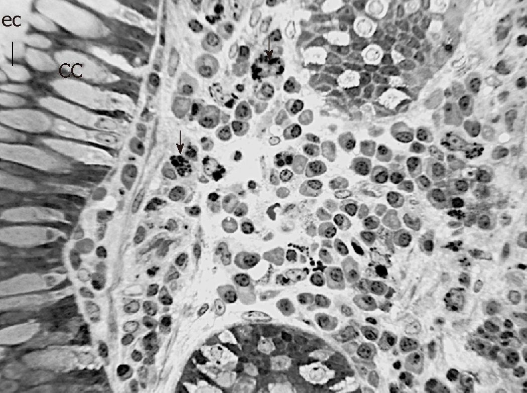
Figure 2 Representative Light microscope (LM) of the lamina propria between Lieberkühn crypts in inflamed ileal tissue of CD patient number 7b.
Notice muciparous goblet cells in cryptal epithelium and large amount and variety of immune cells (arrows) in the connective tissue. Semithin (1 µm thick) section, toluidine blue staining; cc = goblet cell, ec = enterocyte. Scale bar (4 cm) = 70 µm.
- Citation: Giudici F, Lombardelli L, Russo E, Cavalli T, Zambonin D, Logiodice F, Kullolli O, Giusti L, Bargellini T, Fazi M, Biancone L, Scaringi S, Clemente AM, Perissi E, Delfino G, Torcia MG, Ficari F, Tonelli F, Piccinni MP, Malentacchi C. Multiplex gene expression profile in inflamed mucosa of patients with Crohn’s disease ileal localization: A pilot study. World J Clin Cases 2019; 7(17): 2463-2476
- URL: https://www.wjgnet.com/2307-8960/full/v7/i17/2463.htm
- DOI: https://dx.doi.org/10.12998/wjcc.v7.i17.2463


