Copyright
©The Author(s) 2019.
World J Clin Cases. Sep 6, 2019; 7(17): 2413-2419
Published online Sep 6, 2019. doi: 10.12998/wjcc.v7.i17.2413
Published online Sep 6, 2019. doi: 10.12998/wjcc.v7.i17.2413
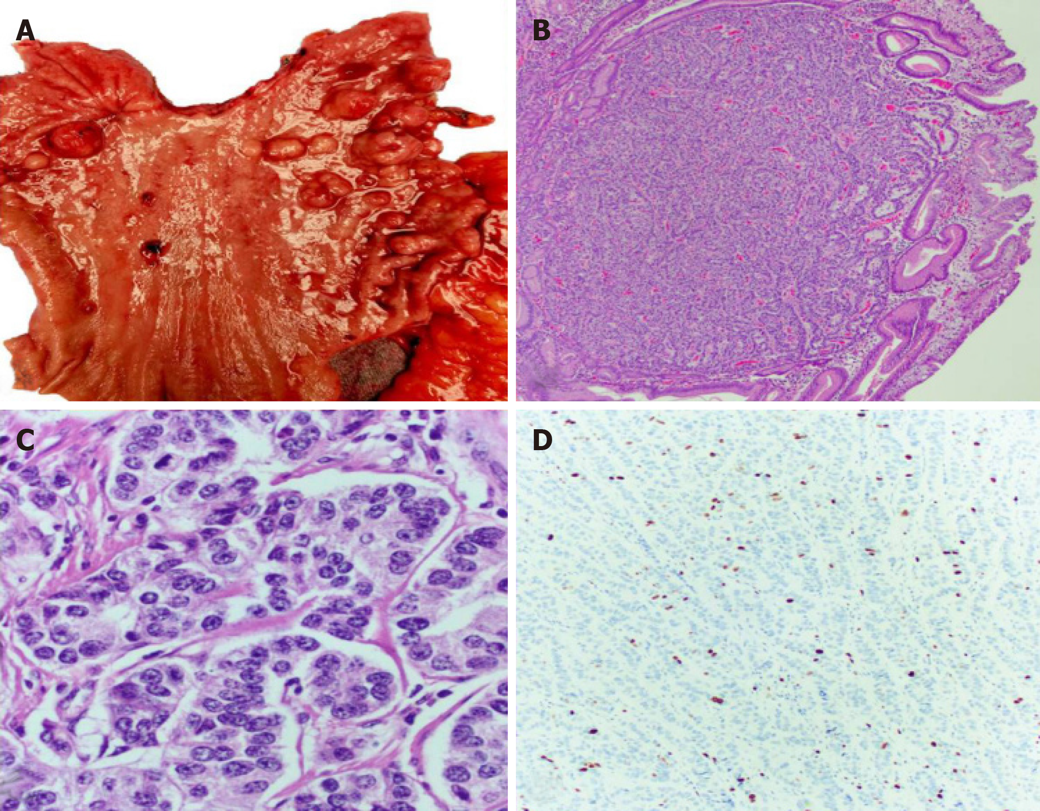
Figure 1 The neuroendocrine tumors are seen as minute (less than 1-2 cm) and frequently multiple polyps.
A: Gastrectomy specimen with multiple nodules/polyps, ranging in size from 0.5 mm to 2 cm; B: Gastric mucosa showing a neuroendocrine tumor (HE, 40×); C: Nested groups of cells with nuclei showing “salt and pepper” chromatin and no mitotic activity (HE, 400×); D: Ki67 proliferation index of less than 3%, consistent with a well differentiated neuroendocrine tumor G1 (Ki67 immunostain, 200×).
- Citation: Algashaamy K, Garcia-Buitrago M. Multifocal G1-G2 gastric neuroendocrine tumors: Differentiating between Type I, II and III, a clinicopathologic review. World J Clin Cases 2019; 7(17): 2413-2419
- URL: https://www.wjgnet.com/2307-8960/full/v7/i17/2413.htm
- DOI: https://dx.doi.org/10.12998/wjcc.v7.i17.2413


