Published online Sep 6, 2019. doi: 10.12998/wjcc.v7.i17.2536
Peer-review started: May 10, 2019
First decision: June 21, 2019
Revised: June 22, 2019
Accepted: July 20, 2019
Article in press: July 20, 2019
Published online: September 6, 2019
Colo-colonic intussusception is an uncommon phenomenon in an adult. Adult intussusception accounts for < 5% of total cases, and the colo-colonic type is < 30% of cases. Although surgical management has been the treatment of choice for intestinal intussusception in adults, because most frequent causes for adult intussusception are malignant in origin, the importance of the roles of preoperative colonoscopic evaluation has recently been emerging.
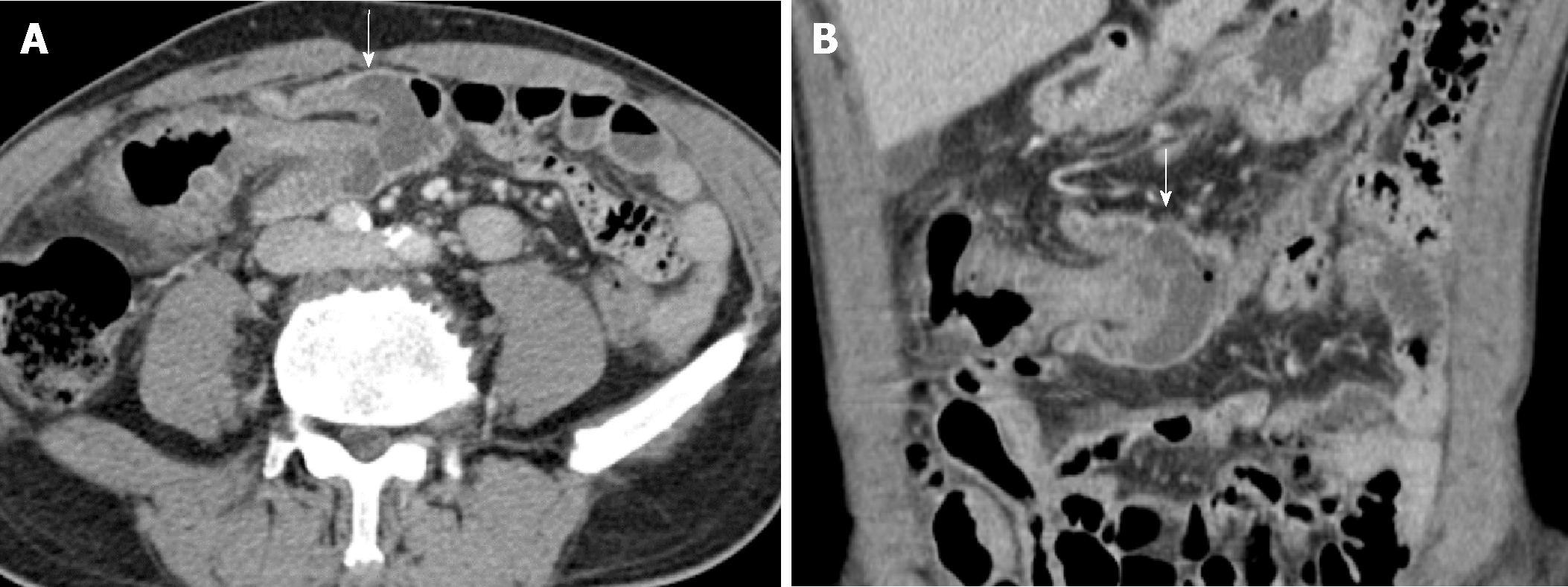
We report an extremely rare case of adult colo-colonic intussusception caused by colonic anisakiasis and successfully treated by endoscopic removal of the Anisakis body. A 59-year-old man visited the emergency department due to 1 day of lower abdominal colicky pain. Abdominopelvic computed tomography (APCT) revealed the presence of mid-transverse colon intussusception without definite necrosis, which was possibly related with colorectal cancer. Because there was no evidence of necrosis at the intussusception site, a colonoscopy was performed to target the colonic lesion and obtain tissue for a histopathological diagnosis. An Anisakis body was found when inspecting the suspicious colonic lesion recorded by APCT. The Anisakis body was removed with forceps assisted by colonoscopy. The patient’s symptoms improved dramatically after removing the Anisakis. A reduced colon without any pathological findings was seen on the follow-up APCT. Without any further treatment, the patient was discharged 5 d after the endoscopy.
When colonic intussusception without necrosis occurs in an adult, physician should consider a colonoscopy to exclude causes cured by endoscopy.
Core tip: We report an extremely rare case of adult colo-colonic intussusception caused by colonic anisakiasis and successfully treated by endoscopic removal of the Anisakis body. When colonic intussusception occurs in an adult patient and there is no evidence of necrosis of the colon, it is necessary to consider a colonoscopy to exclude causes that can be cured by endoscopy.
- Citation: Choi YI, Park DK, Cho HY, Choi SJ, Chung JW, Kim KO, Kwon KA, Kim YJ. Adult intussusception caused by colonic anisakis: A case report. World J Clin Cases 2019; 7(17): 2536-2541
- URL: https://www.wjgnet.com/2307-8960/full/v7/i17/2536.htm
- DOI: https://dx.doi.org/10.12998/wjcc.v7.i17.2536
Eating raw marine food is popular not only in Asian countries but also in Europe and the United States, which has allowed parasitic diseases, such as anisakiasis, to emerge worldwide[1-3].
Anisakiasis is a parasitic disorder that occurs after ingesting the larval stages of ascaridoid nematodes[4]. Anisakids use aquatic mammals as their definitive hosts, while humans are the incidental host after eating raw marine food infected with Anisakis third-stage larvae[4,5]. The disease of anisakiasis presents after direct penetration of larvae into the gastrointestinal wall (invasive) and/or an allergic reaction (noninvasive form)[6]. The invasive form of Anisakis larvae is usually found in the mucosa or submucosa of the gastric and small bowel walls, and causes problems, such as nausea, vomiting, hematemesis, and abdominal pain within a few hours after ingesting the larvae[6]. Colonic anisakiasis is rare, and induces symptoms that mimic other diseases, such as acute appendicitis, ileitis, diverticulitis, cholecystitis, inflammatory bowel disease, and small bowel obstruction[7-10].
Colo-colonic intussusception is also an uncommon phenomenon in an adult[11]. Adult intussusception accounts for < 5% of total cases, and the colo-colonic type is < 30% of cases, while the intestinal type accounts for most of the cases[11].
We report an extremely rare case of adult colo-colonic intussusception caused by anisakiasis, which was successfully treated by a colonoscopic intervention.
A 59-year-old male visited the emergency department presenting with new onset left-sided lower abdominal pain.
He has abdominal pain for 1 day with no febrile sense.
He has no known medical history.
He had no specific personal or family history of cancer or cancer related disease.
Physical examination on admission revealed that he has upper abdominal tenderness but without rebound tenderness.
Laboratory findings, including a blood cell count, chemistry, electrolytes, C-reactive protein (CRP), and carcinoembryonic antigen (CEA) levels were within normal ranges (hemoglobin, 12.9 g/dL; white blood cell count, 7.04 × 10³/mm3; CRP, 0.04 mg/dL; and CEA, 0.69 mg/dL).
He underwent a contrast enhanced abdominopelvic computed tomography scan (APCT) that showed colo-colonic intussusception in the mid-transverse colon (Figure 1). After an interdisciplinary approach on the part of the radiology, surgery, and gastroenterology departments, intussusception induced by descending colon cancer was highly suspected even though there was no definite evidence of cancer on the APCT, given that the most frequent etiology for adult colo-colonic intussusception is malignancy.
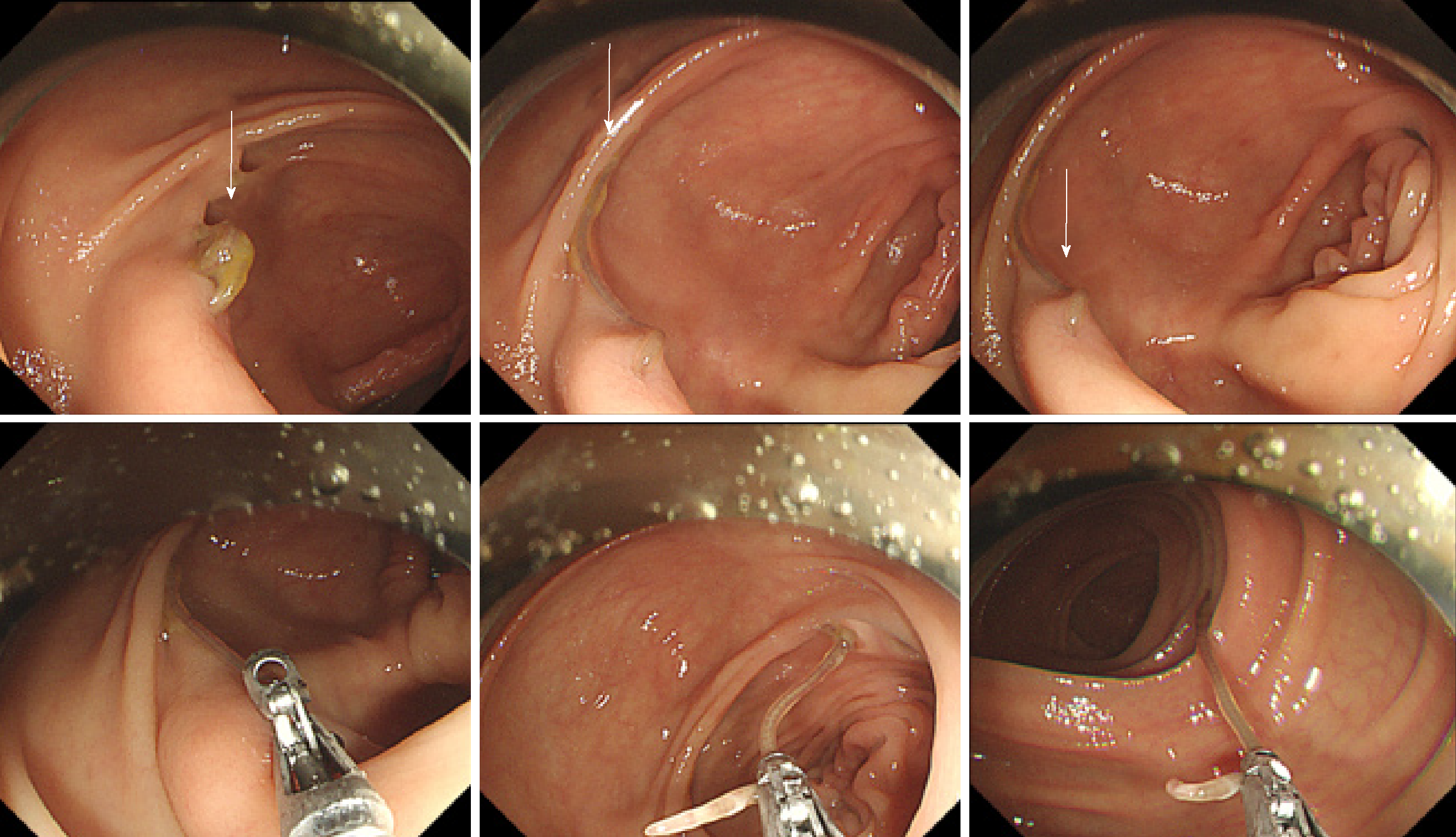
A multidisciplinary team decided on surgery to reduce the intussusception and treat the malignancy. Before the operation, a colonoscopy was done to target the unrevealed malignancy, as there was no definite necrosis around the colonic region with intussusception. During colonoscopic procedures, An approximately 3.0 cm sized Anisakis body, which had penetrated the colonic wall was seen in the transverse colon. The final diagnosis for patient was colo-colonic intussusception caused by colonic anisakiasis.
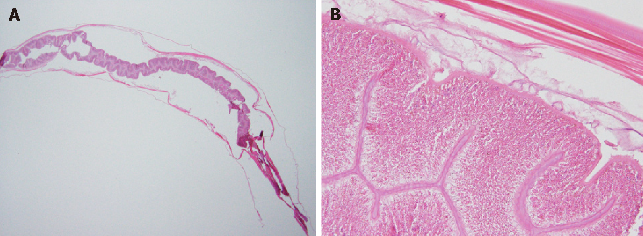
During colonoscopy, an approximately 3.0 cm sized Anisakis body, which had penetrated the colonic wall was seen in the transverse colon (Figure 2). The Anisakis body was removed with biopsy forceps and carried to the pathology department. The final tentative diagnosis was colonic Anisakis (Figure 3).
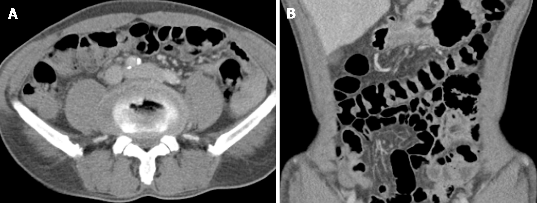
After removing the Anisakis body by colonoscopy, the patient’s symptoms and lower abdominal pain completely resolved and did not require further intervention. A subsequent APCT showed successful reduction of the endoscopic intussusception (Figure 4). Without any further treatment, the patient was discharged 5 days after the endoscopy. After 6month follow up, all of the patient symptom and signs have been alleviated.
In this study, we report an extremely rare case of colo-colonic intussusception in an adult that occurred due to colonic Anisakis and was cured by endoscopic removal of Anisakis larvae.
Adult intestinal intussusception is a rare disease, which accounts for only 5% of all cases of intussusception[12]. Furthermore, colo-colonic intussusception is rarer than small bowel intussusception, and accounts for < 30% of cases[12]. The most common site for colo-colonic intussusception is the proximal colon, and colo-colonic intussusception in the transverse colon is particularly rare [11].
Cases of intestinal intussusception in adults and children differ in a number of aspects, including incidence, etiology, and treatment[12]. While child intussusception includes almost 95% of all intussusception cases, adult intussusception accounts for < 5% of all cases. It is the most common cause of obstruction in infants but is extremely rare in adult and accounts for < 1% of cases. While child intussusception is usually of the primary type, adult intussusception is always secondary to a benign or malignant intestinal neoplasm, stricture, or diverticulum[11]. Intussusception in children is usually treated with supportive care or a contrast enema, whereas 70–90% of adult intussusceptions often require definitive treatment and surgical resection as adult intussusception is generally secondary to a benign or malignant neoplasm, diverticulum, or stricture. In this case, a rare colo-colonic intussusception occurred caused by colonic anisakiasis[13].
Intestinal anisakiasis is a parasitic disease of the gastrointestinal tract. Nematodes in the Anisakidae family cause anisakiasis[14]. Humans are the incidental host in the life cycle after eating raw or uncooked fish containing Anisakis larvae[14]. In this regard, intestinal anisakiasis is prevalent in Asian countries where sushi or raw fish is popular.
The mechanism that precipitates intestinal intussusception is unclear. It has generally been accepted that an injury or irritation of the intestinal wall can alter normal peristalsis of the intestine related with the process of invagination[13]. Colonic Anisakis is a cause of wall injury, particularly when the Anisakis larva penetrates the colon wall; therefore, colonic Anisakis is a cause of colo-colonic intussusception even though colonic Anisakis is extremely rare. In this case, colonic Anisakis may have been the cause of the colo-colonic intussusception, as the patient’s symptoms were relieved and the colonic intussusception was reduced after removing the larva[7,13].
In contrast to previously reported cases, colo-colonic intussusception in this case was successfully resolved by endoscopic reduction in an acute setting (< 24 h), and the cause of the intussusception was promptly diagnosed. Surgical management has been the treatment of choice for intestinal intussusception in adults, because most frequent causes for adult intussusception are malignant in origin. However, a preoperative colonoscopy should be carefully considered especially for cases with no evidence of necrosis of colon in initial imaging study to detect the cause which may be amenable to endoscopic intervention and to avoid an unnecessary surgical procedure. In this case, colo-colonic intussusception, without any evidence of colonic necrosis on the initial image studies including abdominal pelvic computed tomography, was successfully resolved by endoscopic reduction in an acute setting (< 24 h). Further studies comparing endoscopic (or preoperative diagnostic endoscopy) and surgical procedures on long-term outcomes are needed.
The treatment of choice for anisakiasis is endoscopic removal of the nematode, and the clinical symptoms usually stop[15]. In this case, the patient’s symptoms were relieved after endoscopic removal of the Anisakis larva[15]. However, chronic cases with the evidence of the infected lesions on intestine should be considered surgical resection, and administration of tribendazole even though it is ineffective[16].
In conclusion, when colo-colonic intussusception occurs in adult patients, and without definite evidence of necrosis in the intestinal tract, a colonoscopy should be considered to exclude the cause that could be cured by endoscopy and to avoid an unnecessary surgical procedure. Further studies comparing endoscopic (or preoperative diagnostic endoscopy) and surgical procedures on long-term outcomes are needed.
CARE Checklist statement: The authors have read the CARE Checklist and the manuscript was prepared and revised according to the CARE Checklist.
Manuscript source: Unsolicited Manuscript
Specialty type: Medicine, research and experimental
Country of origin: South Korea
Peer-review report classification
Grade A (Excellent): 0
Grade B (Very good): B
Grade C (Good): 0
Grade D (Fair): 0
Grade E (Poor): 0
P-Reviewer: Ziogas DE S-Editor: Cui LJ L-Editor: A E-Editor: Zhou BX
| 1. | Bao M, Pierce GJ, Pascual S, González-Muñoz M, Mattiucci S, Mladineo I, Cipriani P, Bušelić I, Strachan NJ. Assessing the risk of an emerging zoonosis of worldwide concern: anisakiasis. Sci Rep. 2017;7:43699. [PubMed] [DOI] [Cited in This Article: ] [Cited by in Crossref: 136] [Cited by in F6Publishing: 103] [Article Influence: 14.7] [Reference Citation Analysis (0)] |
| 2. | Bucci C, Gallotta S, Morra I, Fortunato A, Ciacci C, Iovino P. Anisakis, just think about it in an emergency! Int J Infect Dis. 2013;17:e1071-e1072. [PubMed] [DOI] [Cited in This Article: ] [Cited by in Crossref: 31] [Cited by in F6Publishing: 28] [Article Influence: 2.5] [Reference Citation Analysis (0)] |
| 3. | Garcia-Perez JC, Rodríguez-Perez R, Ballestero A, Zuloaga J, Fernandez-Puntero B, Arias-Díaz J, Caballero ML. Previous Exposure to the Fish Parasite Anisakis as a Potential Risk Factor for Gastric or Colon Adenocarcinoma. Medicine (Baltimore). 2015;94:e1699. [PubMed] [DOI] [Cited in This Article: ] [Cited by in Crossref: 31] [Cited by in F6Publishing: 30] [Article Influence: 3.3] [Reference Citation Analysis (0)] |
| 4. | Cavallero S, Martini A, Migliara G, De Vito C, Iavicoli S, D'Amelio S. Anisakiasis in Italy: Analysis of hospital discharge records in the years 2005-2015. PLoS One. 2018;13:e0208772. [PubMed] [DOI] [Cited in This Article: ] [Cited by in Crossref: 13] [Cited by in F6Publishing: 16] [Article Influence: 2.7] [Reference Citation Analysis (0)] |
| 5. | Yera H, Fréalle É, Dutoit E, Dupouy-Camet J. A national retrospective survey of anisakidosis in France (2010-2014): decreasing incidence, female predominance, and emerging allergic potential. Parasite. 2018;25:23. [PubMed] [DOI] [Cited in This Article: ] [Cited by in Crossref: 18] [Cited by in F6Publishing: 20] [Article Influence: 3.3] [Reference Citation Analysis (0)] |
| 6. | Sohn WM, Na BK, Kim TH, Park TJ. Anisakiasis: Report of 15 Gastric Cases Caused by Anisakis Type I Larvae and a Brief Review of Korean Anisakiasis Cases. Korean J Parasitol. 2015;53:465-470. [PubMed] [DOI] [Cited in This Article: ] [Cited by in Crossref: 35] [Cited by in F6Publishing: 36] [Article Influence: 4.0] [Reference Citation Analysis (0)] |
| 7. | Tamai Y, Kobayashi K. Asymptomatic colonic anisakiasis. Intern Med. 2015;54:675. [PubMed] [DOI] [Cited in This Article: ] [Cited by in Crossref: 8] [Cited by in F6Publishing: 9] [Article Influence: 1.0] [Reference Citation Analysis (0)] |
| 8. | Schuster R, Petrini JL, Choi R. Anisakiasis of the colon presenting as bowel obstruction. Am Surg. 2003;69:350-352. [PubMed] [Cited in This Article: ] |
| 9. | Yoo HJ, Kim SH, Lee JM, Kim MA, Han JK, Choi BI. The association of anisakiasis in the ascending colon with sigmoid colon cancer: CT colonography findings. Korean J Radiol. 2008;9 Suppl:S56-S60. [PubMed] [DOI] [Cited in This Article: ] [Cited by in Crossref: 15] [Cited by in F6Publishing: 15] [Article Influence: 0.9] [Reference Citation Analysis (0)] |
| 10. | Yorimitsu N, Hiraoka A, Utsunomiya H, Imai Y, Tatsukawa H, Tazuya N, Yamago H, Shimizu Y, Hidaka S, Tanihira T, Hasebe A, Miyamoto Y, Ninomiya T, Abe M, Hiasa Y, Matsuura B, Onji M, Michitaka K. Colonic intussusception caused by anisakiasis: a case report and review of the literature. Intern Med. 2013;52:223-226. [PubMed] [Cited in This Article: ] |
| 11. | Wang N, Cui XY, Liu Y, Long J, Xu YH, Guo RX, Guo KJ. Adult intussusception: a retrospective review of 41 cases. World J Gastroenterol. 2009;15:3303-3308. [PubMed] [DOI] [Cited in This Article: ] [Cited by in Crossref: 140] [Cited by in F6Publishing: 1] [Article Influence: 0.1] [Reference Citation Analysis (0)] |
| 12. | Honjo H, Mike M, Kusanagi H, Kano N. Adult intussusception: a retrospective review. World J Surg. 2015;39:134-138. [PubMed] [DOI] [Cited in This Article: ] [Cited by in Crossref: 118] [Cited by in F6Publishing: 143] [Article Influence: 15.9] [Reference Citation Analysis (0)] |
| 13. | Ramos L, Alonso C, Guilarte M, Vilaseca J, Santos J, Malagelada JR. Anisakis simplex-induced small bowel obstruction after fish ingestion: preliminary evidence for response to parenteral corticosteroids. Clin Gastroenterol Hepatol. 2005;3:667-671. [PubMed] [DOI] [Cited in This Article: ] [Cited by in Crossref: 20] [Cited by in F6Publishing: 20] [Article Influence: 1.1] [Reference Citation Analysis (0)] |
| 14. | Mumoli N, Merlo A. Colonic anisakiasis. CMAJ. 2013;185:E652. [PubMed] [DOI] [Cited in This Article: ] [Cited by in Crossref: 6] [Cited by in F6Publishing: 6] [Article Influence: 0.5] [Reference Citation Analysis (0)] |
| 15. | Kojima G, Usuki S, Mizokami K, Tanabe M, Machi J. Intestinal anisakiasis as a rare cause of small bowel obstruction. Am J Emerg Med. 2013;31:1422.e1-1422.e2. [PubMed] [DOI] [Cited in This Article: ] [Cited by in Crossref: 15] [Cited by in F6Publishing: 17] [Article Influence: 1.5] [Reference Citation Analysis (0)] |
| 16. | Guardone L, Armani A, Nucera D, Costanzo F, Mattiucci S, Bruschi F. Human anisakiasis in Italy: a retrospective epidemiological study over two decades. Parasite. 2018;25:41. [PubMed] [DOI] [Cited in This Article: ] [Cited by in Crossref: 57] [Cited by in F6Publishing: 55] [Article Influence: 9.2] [Reference Citation Analysis (0)] |


