Copyright
©The Author(s) 2019.
World J Clin Cases. Aug 26, 2019; 7(16): 2406-2412
Published online Aug 26, 2019. doi: 10.12998/wjcc.v7.i16.2406
Published online Aug 26, 2019. doi: 10.12998/wjcc.v7.i16.2406
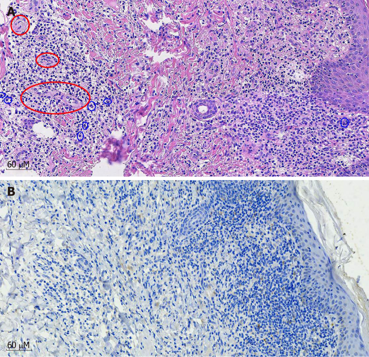
Figure 3 Photomicrograph of section of the skin biopsy from the abdomen lesion.
A: The mixed infiltrate of lymphocytes, histocytes, and plasma cells (blue circle) accompanied by obliterative vasculitis in the dermis (red circle). (Hematoxylin-eosin staining, original magnification, ×200); B: Spiral and thread-like organisms, highlighted by the brown chromogen in the dermis, represent the spirochetes. (Immunohistochemical staining with anti-spirochetes, original magnification, ×200).
- Citation: Ge G, Li DM, Qiu Y, Fu HJ, Zhang XY, Shi DM. Malignant syphilis accompanied with neurosyphilis in a malnourished patient: A case report. World J Clin Cases 2019; 7(16): 2406-2412
- URL: https://www.wjgnet.com/2307-8960/full/v7/i16/2406.htm
- DOI: https://dx.doi.org/10.12998/wjcc.v7.i16.2406


