Copyright
©The Author(s) 2019.
World J Clin Cases. Aug 26, 2019; 7(16): 2393-2400
Published online Aug 26, 2019. doi: 10.12998/wjcc.v7.i16.2393
Published online Aug 26, 2019. doi: 10.12998/wjcc.v7.i16.2393
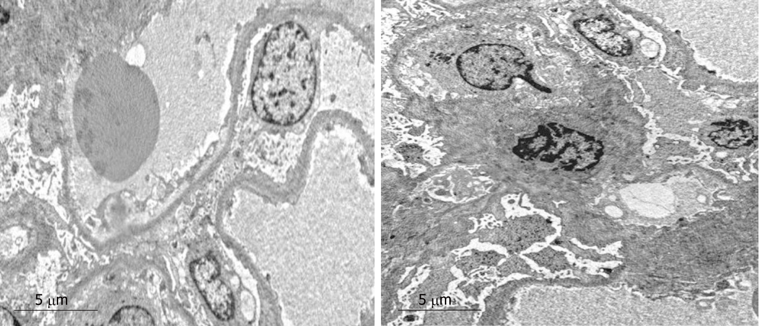
Figure 3 Electron microscopy.
Extensive effacement of podocyte foot processes, slight hyperplasia of mesangial matrix, and small amounts of electron dense depositions were observed in the mesangial area. Interstitial fibrosis of the kidney was obvious, and inflammatory cell infiltration was seen.
- Citation: Mwamunyi MJ, Zhu HY, Zhang C, Yuan YP, Yao LJ. Pseudothrombus deposition accompanied with minimal change nephrotic syndrome and chronic kidney disease in a patient with Waldenström's macroglobulinemia: A case report. World J Clin Cases 2019; 7(16): 2393-2400
- URL: https://www.wjgnet.com/2307-8960/full/v7/i16/2393.htm
- DOI: https://dx.doi.org/10.12998/wjcc.v7.i16.2393


