Copyright
©The Author(s) 2019.
World J Clin Cases. Aug 26, 2019; 7(16): 2393-2400
Published online Aug 26, 2019. doi: 10.12998/wjcc.v7.i16.2393
Published online Aug 26, 2019. doi: 10.12998/wjcc.v7.i16.2393
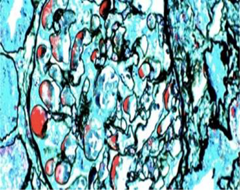
Figure 2 Light microscopy.
Periodic Schiff-Methenamine (PASM) and Masson staining. Fuchsinophilic depositions were found in the basement membrane and under the endothelium. The tubulointerstitium exhibited moderate lesions, with acute lesions on chronic damage. There was diffuse turbidity and granular degeneration in the tubular epithelial cells. Partial tubular epithelial cells presented small and fine vacuolar degeneration, and the basement membrane of tubules became thicker. Brush border of the tubules was absent. Protein casts could be seen in some lumens. The renal interstitial region could be found to be focally enlarged, and fibrosis index was 1+. Individual arterioles presented segmental hyalinosis (PASM and Masson staining; magnification, ×400).
- Citation: Mwamunyi MJ, Zhu HY, Zhang C, Yuan YP, Yao LJ. Pseudothrombus deposition accompanied with minimal change nephrotic syndrome and chronic kidney disease in a patient with Waldenström's macroglobulinemia: A case report. World J Clin Cases 2019; 7(16): 2393-2400
- URL: https://www.wjgnet.com/2307-8960/full/v7/i16/2393.htm
- DOI: https://dx.doi.org/10.12998/wjcc.v7.i16.2393


