Copyright
©The Author(s) 2019.
World J Clin Cases. Aug 26, 2019; 7(16): 2393-2400
Published online Aug 26, 2019. doi: 10.12998/wjcc.v7.i16.2393
Published online Aug 26, 2019. doi: 10.12998/wjcc.v7.i16.2393
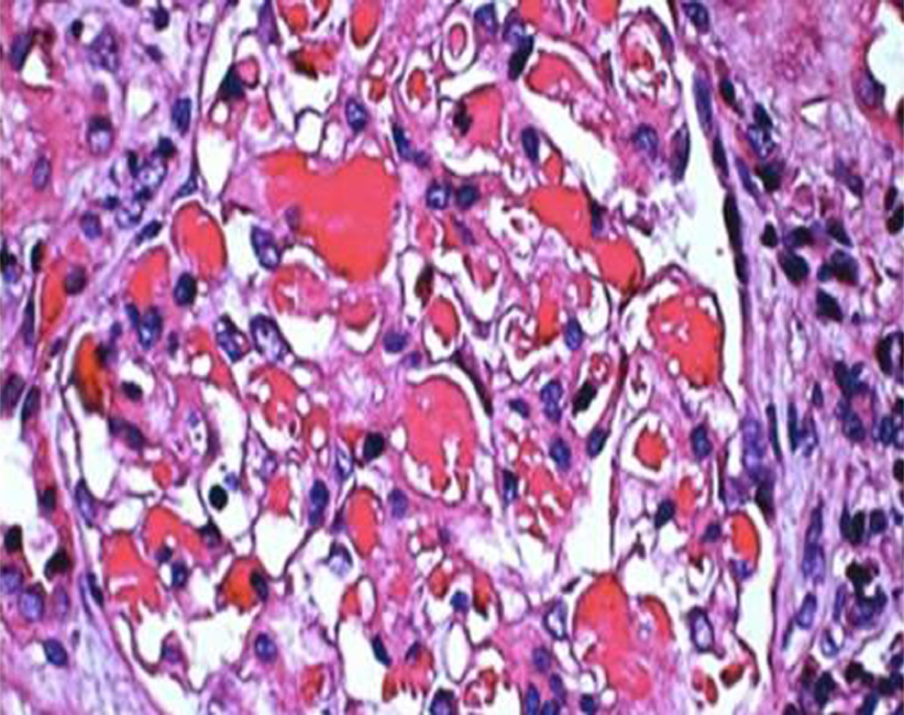
Figure 1 Light microscopy.
The volume of residual glomeruli increased, the number of cells was 80-120 per glomerulus, mesangial cells and mesangial matrix were slightly increased, capillary loops were open, and the number of infiltrating cells was < 3 per glomeruli, mainly mononuclear cells. Red blood cells and “pseudothrombi” could be seen in several capillaries. One capillary loop was embedded into the urinary pole (HE staining; magnification, ×600).
- Citation: Mwamunyi MJ, Zhu HY, Zhang C, Yuan YP, Yao LJ. Pseudothrombus deposition accompanied with minimal change nephrotic syndrome and chronic kidney disease in a patient with Waldenström's macroglobulinemia: A case report. World J Clin Cases 2019; 7(16): 2393-2400
- URL: https://www.wjgnet.com/2307-8960/full/v7/i16/2393.htm
- DOI: https://dx.doi.org/10.12998/wjcc.v7.i16.2393


