Copyright
©The Author(s) 2019.
World J Clin Cases. Aug 26, 2019; 7(16): 2269-2286
Published online Aug 26, 2019. doi: 10.12998/wjcc.v7.i16.2269
Published online Aug 26, 2019. doi: 10.12998/wjcc.v7.i16.2269
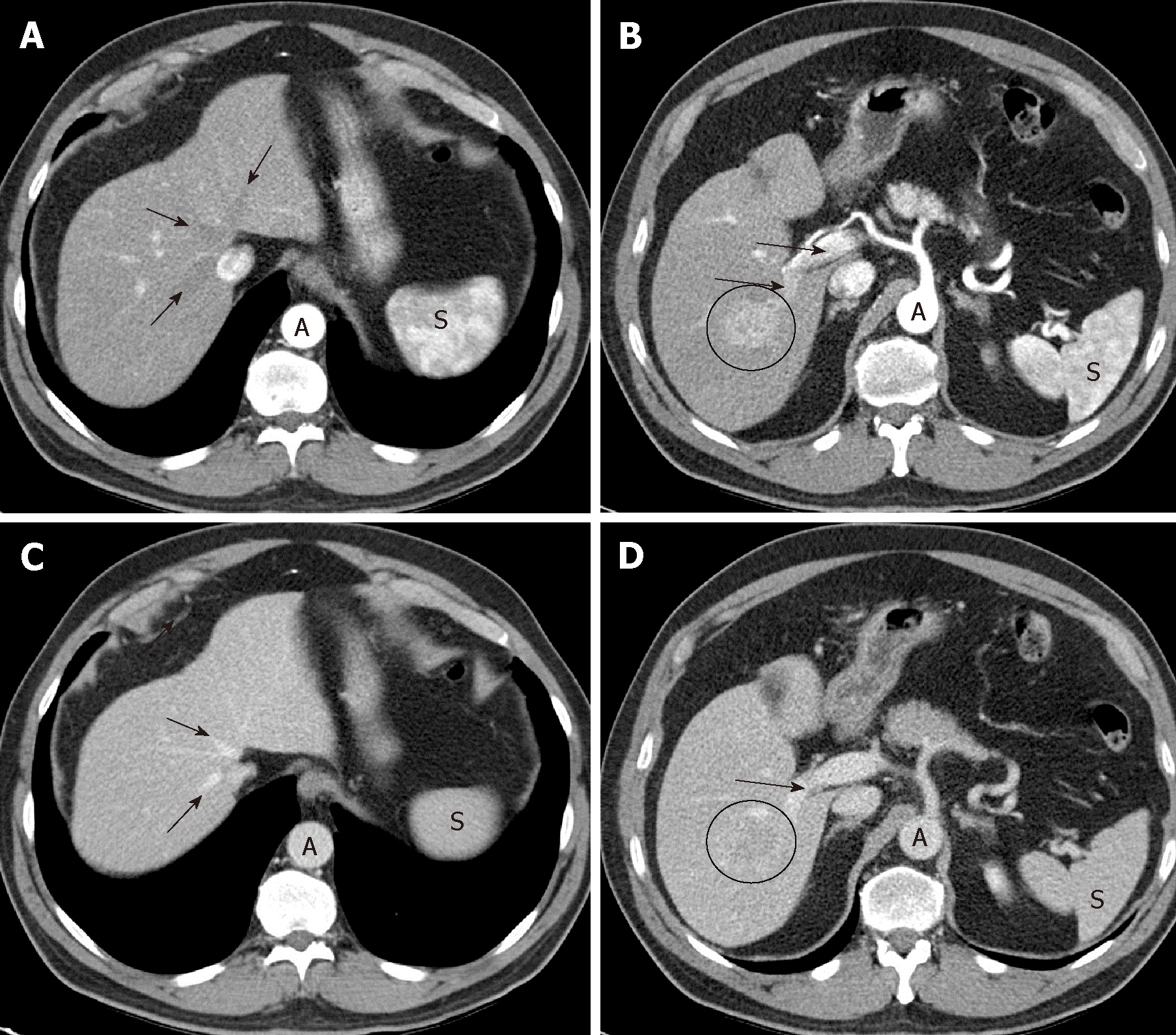
Figure 1 Optimal late arterial phase and portal phase.
A, B: Hepatic artery and branches are fully enhanced. Portal vein is enhanced (arrows) but hepatic veins not yet enhanced by antegrade flow (arrows). Heterogeneous spleen. Aorta of very high density; C, D: Portal phase: portal veins are fully enhanced (D: arrows). Hepatic veins are enhanced by antegrade flow (C: arrows). Liver parenchyma is at peak enhancement. Homogeneous spleen. Portal vein even denser than aorta.
- Citation: Pascual S, Miralles C, Bernabé JM, Irurzun J, Planells M. Surveillance and diagnosis of hepatocellular carcinoma: A systematic review. World J Clin Cases 2019; 7(16): 2269-2286
- URL: https://www.wjgnet.com/2307-8960/full/v7/i16/2269.htm
- DOI: https://dx.doi.org/10.12998/wjcc.v7.i16.2269


