Copyright
©The Author(s) 2019.
World J Clin Cases. Aug 6, 2019; 7(15): 2128-2133
Published online Aug 6, 2019. doi: 10.12998/wjcc.v7.i15.2128
Published online Aug 6, 2019. doi: 10.12998/wjcc.v7.i15.2128
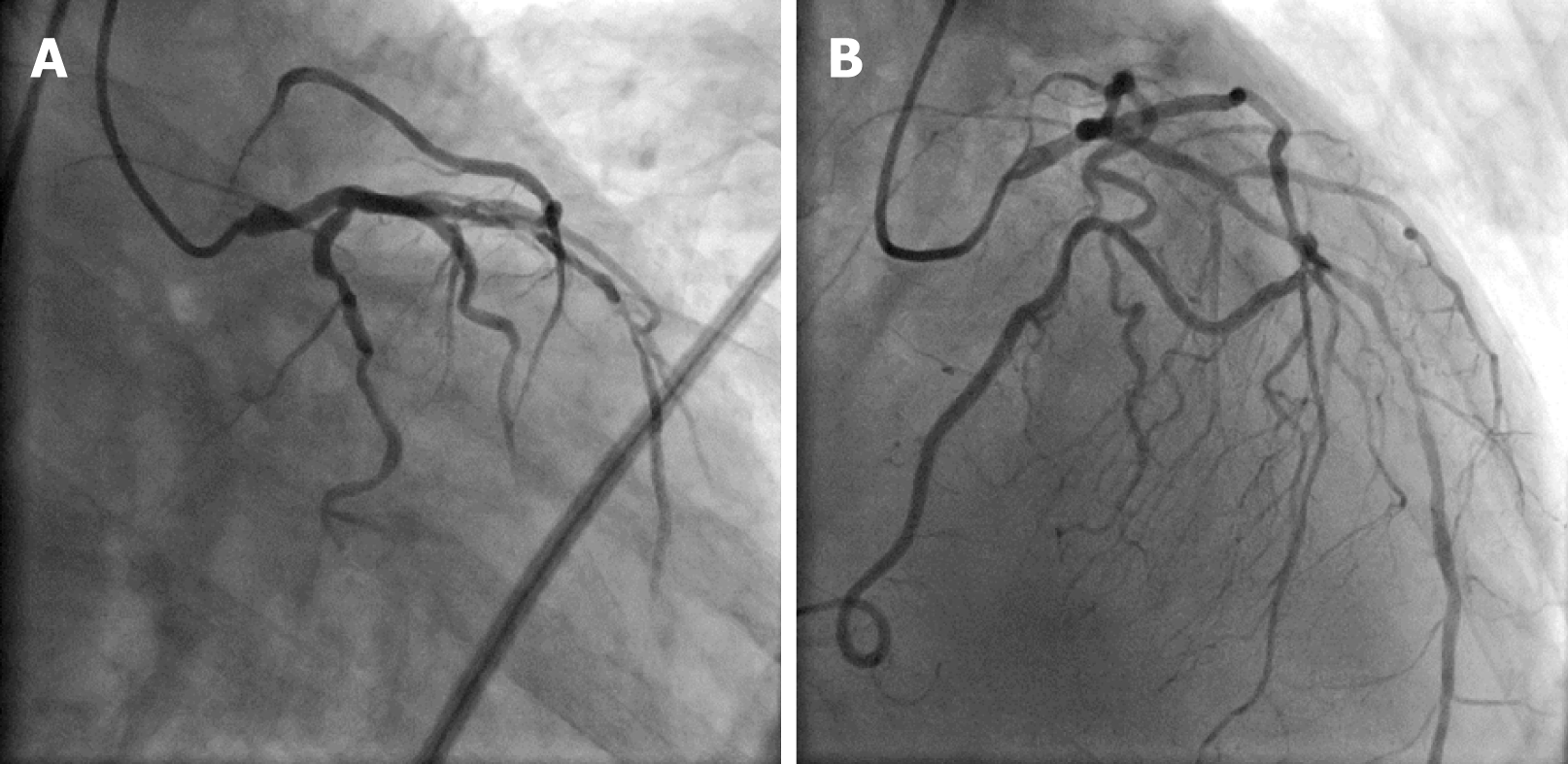
Figure 1 Coronary angiograms.
A: Left anterior oblique caudal view; B: Right anterior oblique cranial view. The coronary angiograms show the left main coronary artery with severe ostial stenosis and left anterior descending artery (LAD) with a diffuse lesion in the mid segment and right coronary artery from the midportion of the LAD.
- Citation: Wu Q, Li ZZ, Yue F, Wei F, Zhang CY. Percutaneous coronary intervention for ostial lesions of the left main stem in a patient with congenital single left coronary artery: A case report. World J Clin Cases 2019; 7(15): 2128-2133
- URL: https://www.wjgnet.com/2307-8960/full/v7/i15/2128.htm
- DOI: https://dx.doi.org/10.12998/wjcc.v7.i15.2128


