Copyright
©The Author(s) 2019.
World J Clin Cases. Aug 6, 2019; 7(15): 2094-2102
Published online Aug 6, 2019. doi: 10.12998/wjcc.v7.i15.2094
Published online Aug 6, 2019. doi: 10.12998/wjcc.v7.i15.2094
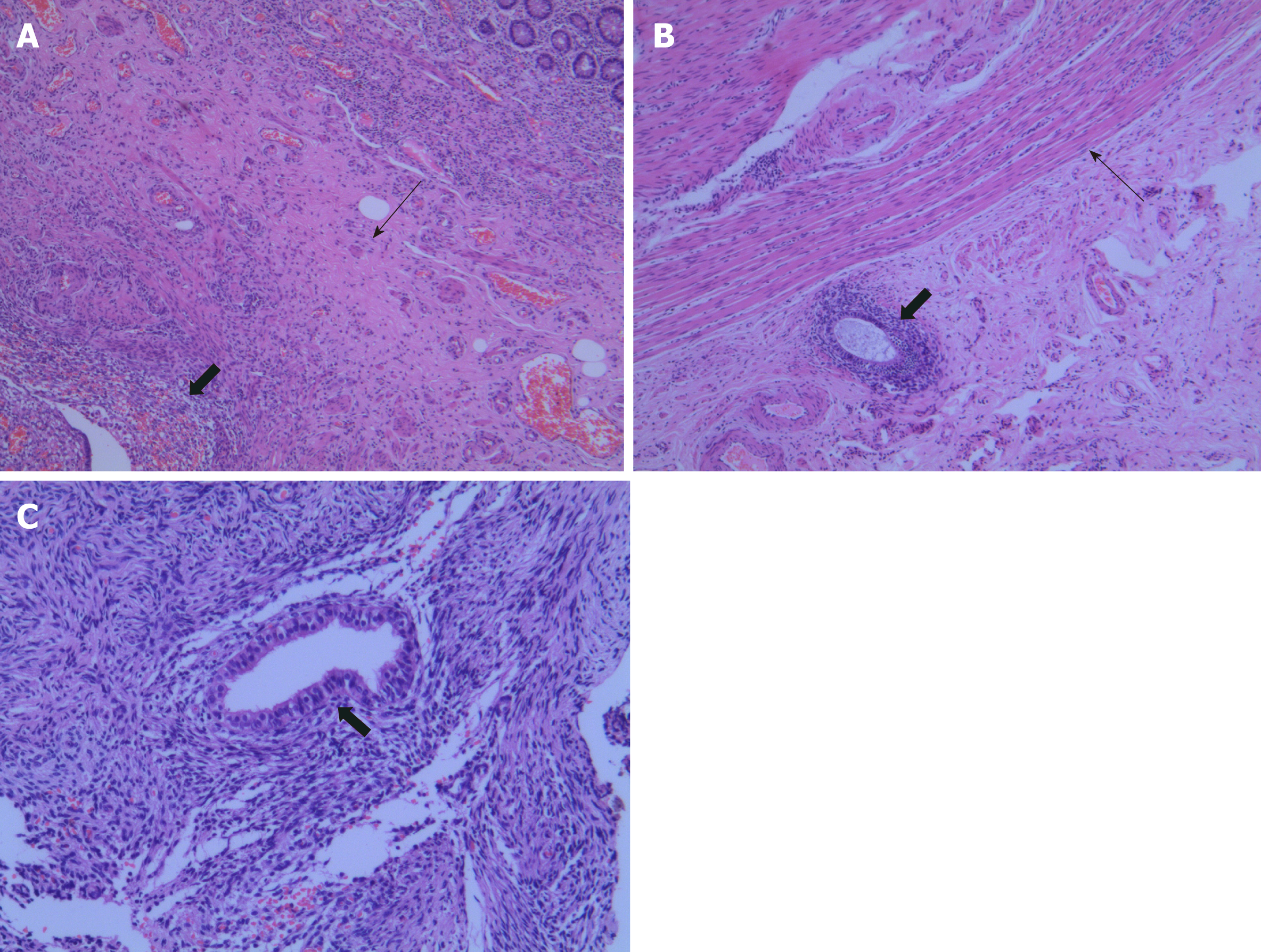
Figure 3 Histopathological appearances of the endometriosis in appendix, small intestine, and ovary.
A: Hematoxylin and eosin (HE) staining shows that ectopic endometrial-type glands and stroma (thick arrows) were detected in the muscular layers (thin arrows) of the appendix (×40); B: Ectopic endometrial-type glands and stroma (thick arrows) were observed in the subserosa of the small intestine. The muscular layers are indicated by thin arrows (HE, ×40); C: Higher magnification of endometriosis (thick arrows) in the ovary (HE, ×100).
- Citation: Zhu MY, Fei FM, Chen J, Zhou ZC, Wu B, Shen YY. Endometriosis of the duplex appendix: A case report and review of the literature. World J Clin Cases 2019; 7(15): 2094-2102
- URL: https://www.wjgnet.com/2307-8960/full/v7/i15/2094.htm
- DOI: https://dx.doi.org/10.12998/wjcc.v7.i15.2094


