Copyright
©The Author(s) 2019.
World J Clin Cases. Jul 26, 2019; 7(14): 1884-1891
Published online Jul 26, 2019. doi: 10.12998/wjcc.v7.i14.1884
Published online Jul 26, 2019. doi: 10.12998/wjcc.v7.i14.1884
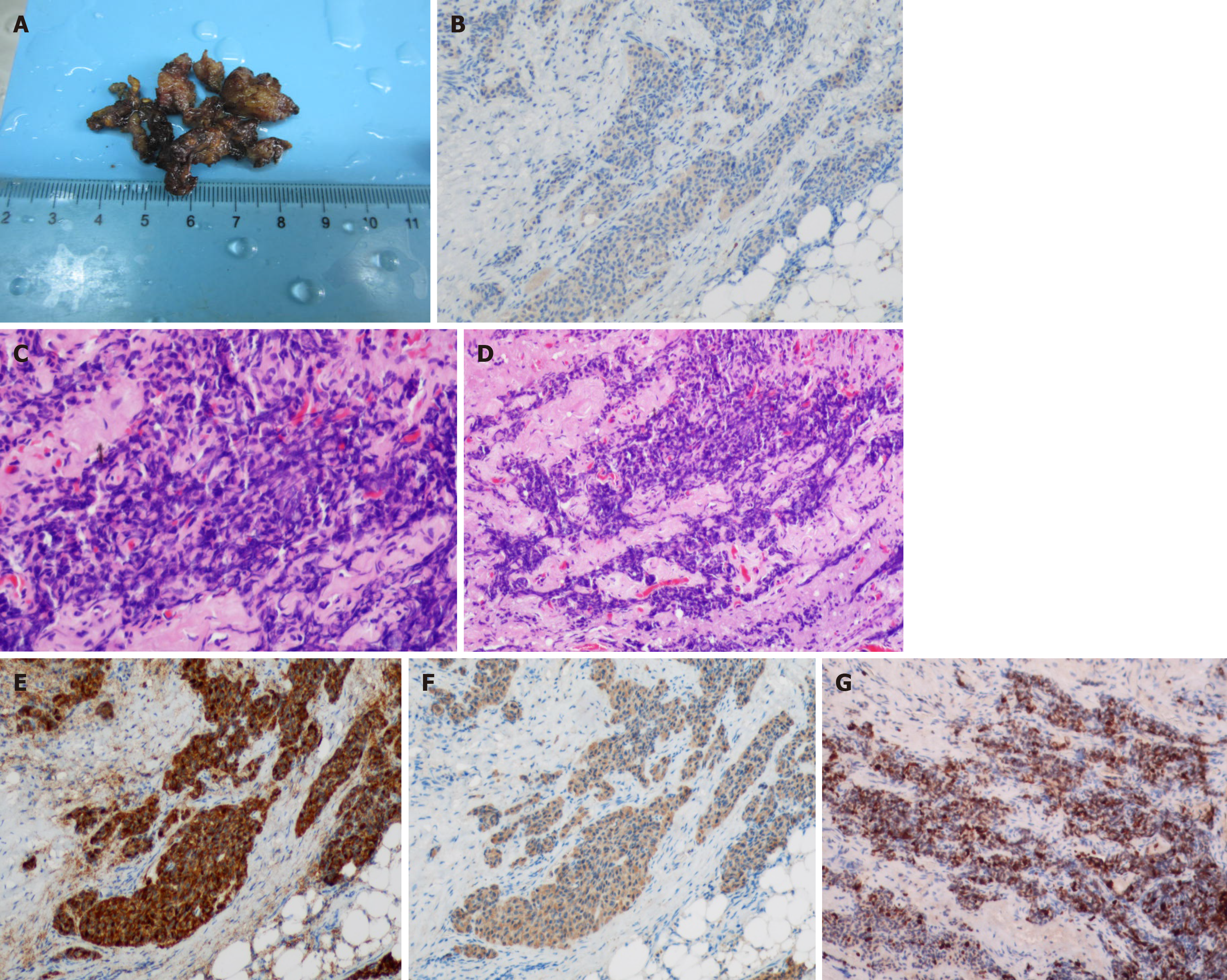
Figure 3 Final pathological images of the tumor.
A: The mass removed by resection; B: The tumor component tested positive for the marker Syn (×100); C: The tumor stained using H&E (×100); D: The tumor stained using H&E (×200); E: The tumor component was diffusely positive for the marker CD56 (×100); F: The tumor component was positive for CK (×100); G: The tumor’s Ki67 index was 30% (×100).
- Citation: Zhang R, Zhu Y, Huang XB, Deng C, Li M, Shen GS, Huang SL, Huangfu SH, Liu YN, Zhou CG, Wang L, Zhang Q, Deng Y, Jiang B. Primary neuroendocrine tumor in the presacral region: A case report. World J Clin Cases 2019; 7(14): 1884-1891
- URL: https://www.wjgnet.com/2307-8960/full/v7/i14/1884.htm
- DOI: https://dx.doi.org/10.12998/wjcc.v7.i14.1884


