Copyright
©The Author(s) 2019.
World J Clin Cases. Jul 26, 2019; 7(14): 1884-1891
Published online Jul 26, 2019. doi: 10.12998/wjcc.v7.i14.1884
Published online Jul 26, 2019. doi: 10.12998/wjcc.v7.i14.1884
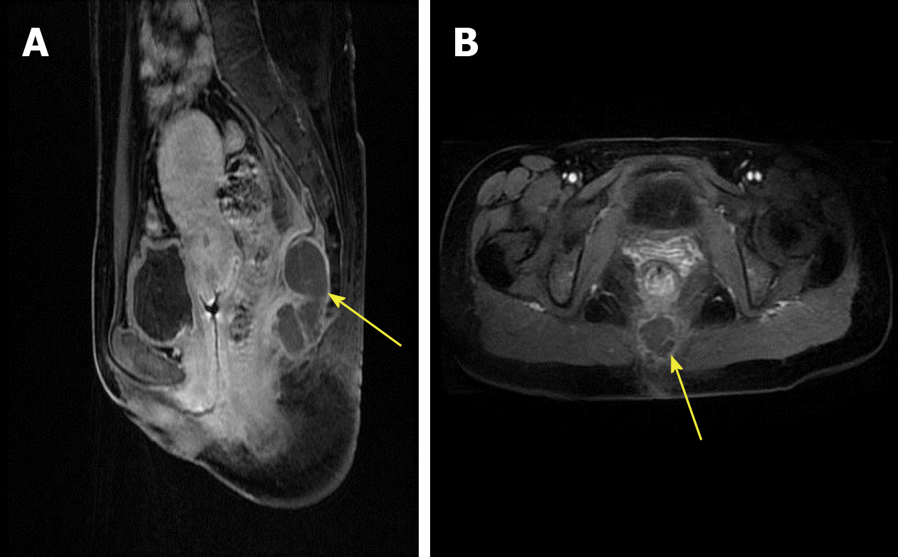
Figure 1 Magnetic resonance imaging scans.
A: Sagittal view demonstrating a 57 mm × 29 mm presacral mass, with margin and separation being visible; B: T2STIR-weighted axial MRI image showing high intensity of the tumor.
- Citation: Zhang R, Zhu Y, Huang XB, Deng C, Li M, Shen GS, Huang SL, Huangfu SH, Liu YN, Zhou CG, Wang L, Zhang Q, Deng Y, Jiang B. Primary neuroendocrine tumor in the presacral region: A case report. World J Clin Cases 2019; 7(14): 1884-1891
- URL: https://www.wjgnet.com/2307-8960/full/v7/i14/1884.htm
- DOI: https://dx.doi.org/10.12998/wjcc.v7.i14.1884


