Copyright
©The Author(s) 2019.
World J Clin Cases. Jul 26, 2019; 7(14): 1865-1875
Published online Jul 26, 2019. doi: 10.12998/wjcc.v7.i14.1865
Published online Jul 26, 2019. doi: 10.12998/wjcc.v7.i14.1865
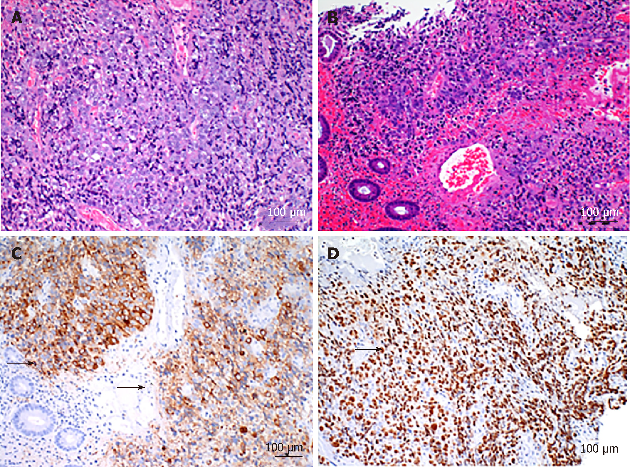
Figure 2 Histological findings of the tumor.
A: Hematoxylin-eosin staining showed that the tumor cells were poorly differentiated; B: Complicated with the intratumoral bleeding; C: Tumor cells underwent immunohistochemical staining for Synaptophysin. Black arrows indicate the positively stained cells; D: A high proliferative fraction of immunohistochemical staining for Ki67 staining. Black arrows indicate the positively stained cells.
- Citation: Yoshida T, Kamimura K, Hosaka K, Doumori K, Oka H, Sato A, Fukuhara Y, Watanabe S, Sato T, Yoshikawa A, Tomidokoro T, Terai S. Colorectal neuroendocrine carcinoma: A case report and review of the literature. World J Clin Cases 2019; 7(14): 1865-1875
- URL: https://www.wjgnet.com/2307-8960/full/v7/i14/1865.htm
- DOI: https://dx.doi.org/10.12998/wjcc.v7.i14.1865


