Copyright
©The Author(s) 2019.
World J Clin Cases. Jul 26, 2019; 7(14): 1857-1864
Published online Jul 26, 2019. doi: 10.12998/wjcc.v7.i14.1857
Published online Jul 26, 2019. doi: 10.12998/wjcc.v7.i14.1857
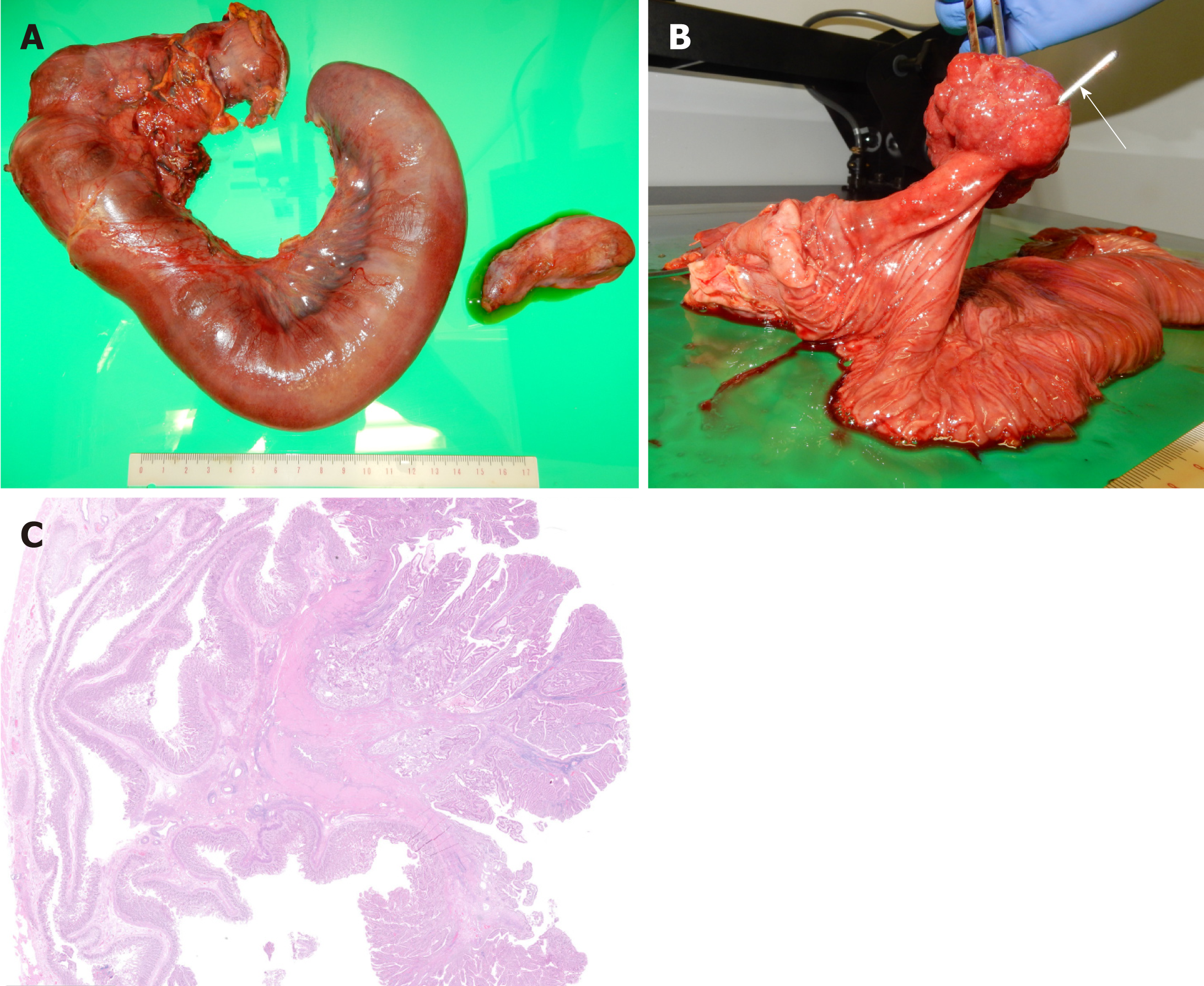
Figure 6 Resected specimen and pathological examination.
A: This image shows a frontal view of the gross pancreaticoduodenectomy specimen; B: The pedunculate ampullary tumor is 4.5 cm × 3.8 cm × 2.6 cm in size. A sonde is passed through the biliary tract (white arrow); C: Histology confirms an ampullary tubulovillous adenoma with no dysplasia originating from the duodenal epithelium in the papilla of Vater. There is no evidence of intraductal extension or malignancy (hematoxylin and eosin staining, × 100).
- Citation: Hirata M, Shirakata Y, Yamanaka K. Duodenal intussusception secondary to ampullary adenoma: A case report. World J Clin Cases 2019; 7(14): 1857-1864
- URL: https://www.wjgnet.com/2307-8960/full/v7/i14/1857.htm
- DOI: https://dx.doi.org/10.12998/wjcc.v7.i14.1857


