Copyright
©The Author(s) 2019.
World J Clin Cases. Jul 26, 2019; 7(14): 1837-1843
Published online Jul 26, 2019. doi: 10.12998/wjcc.v7.i14.1837
Published online Jul 26, 2019. doi: 10.12998/wjcc.v7.i14.1837
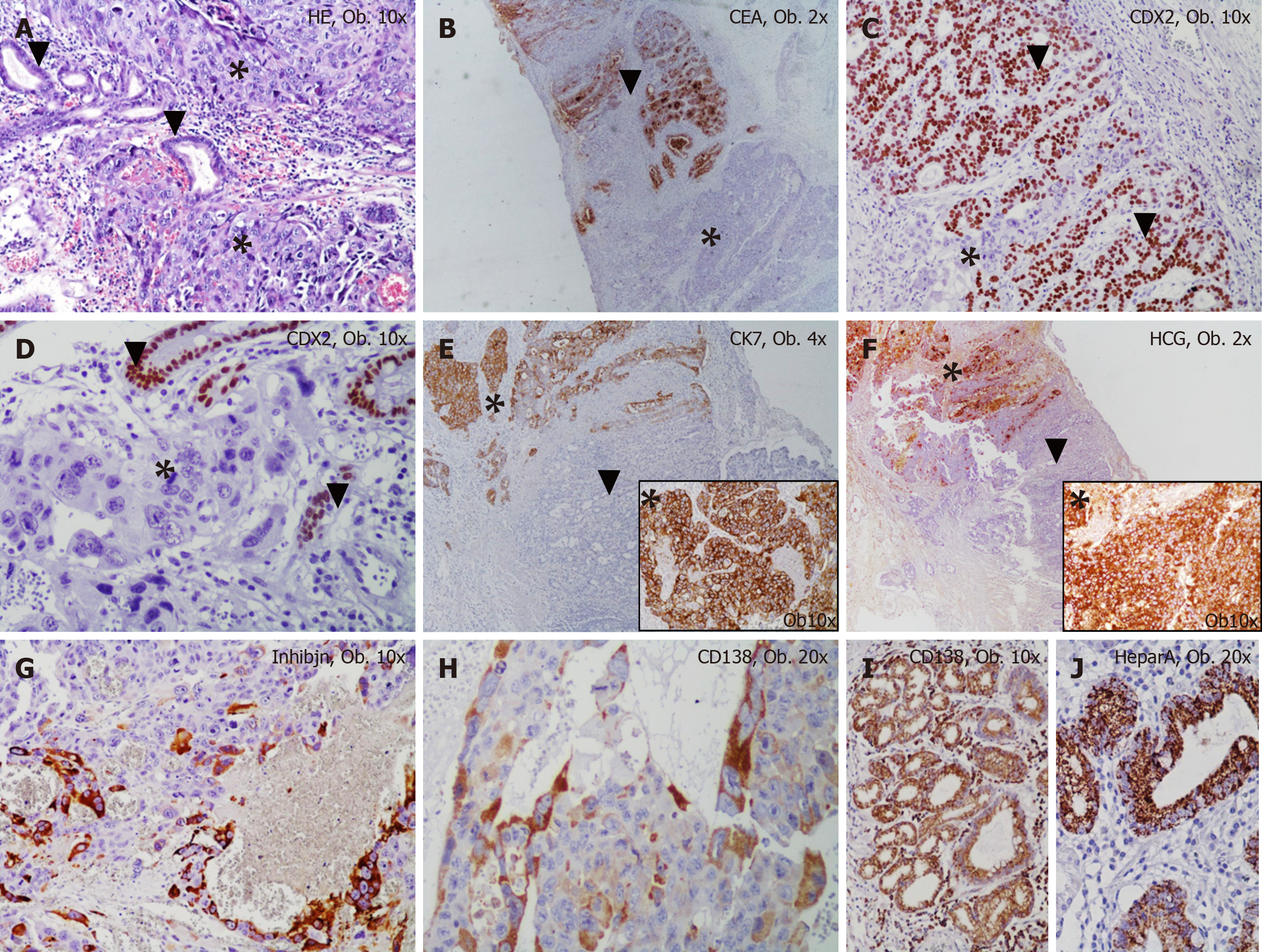
Figure 2 Histological examination results.
A: The mixed gastric carcinoma shows two components: choriocarcinoma (marked by *) and adenocarcinoma (marked by ▼); B-F: The choriocarcinoma cells are negative for CEA (B) and CDX2 (C, D) and show positivity for Cytokeratin 7 (E) and HCG (F); G, H: The syncytiotrophoblast-like cells are marked by inhibin (G) and CD138 (H). The adenocarcinoma component is positive for CEA (B) and CDX2 (C, D) and to not present positivity for Cytokeratin 7 (E) and HCG (F); I, J: The normal gastric mucosa cells are marked by CD138 (I) and Hepar A (J).
- Citation: Gurzu S, Copotoiu C, Tugui A, Kwizera C, Szodorai R, Jung I. Primary gastric choriocarcinoma - a rare and aggressive tumor with multilineage differentiation: A case report. World J Clin Cases 2019; 7(14): 1837-1843
- URL: https://www.wjgnet.com/2307-8960/full/v7/i14/1837.htm
- DOI: https://dx.doi.org/10.12998/wjcc.v7.i14.1837


