Copyright
©The Author(s) 2019.
World J Clin Cases. Jul 26, 2019; 7(14): 1837-1843
Published online Jul 26, 2019. doi: 10.12998/wjcc.v7.i14.1837
Published online Jul 26, 2019. doi: 10.12998/wjcc.v7.i14.1837
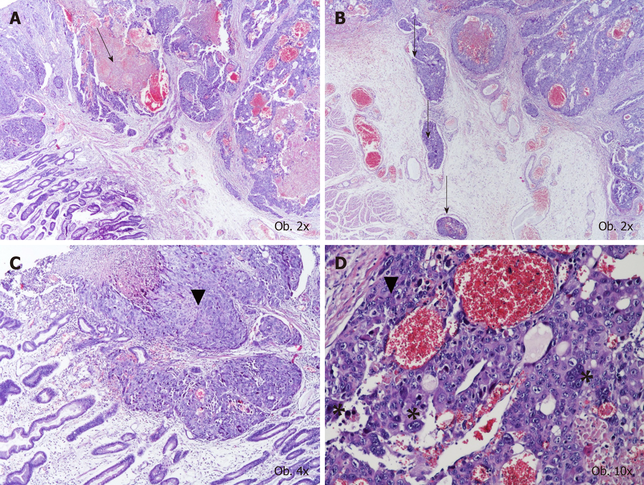
Figure 1 Histological examination results.
A, B: The histological aspect of choriocarcinoma, with large hemorrhagic area (A), multiple vascular emboli (B), and characteristic proliferation of oval cells with pale cytoplasm with similar aspect to cytotrophoblastic cells (marked by ▼) and giant cells with lobulated or bizarre nuclei (marked by *); C, D: Similar to syncytiotrophoblastic cells.
- Citation: Gurzu S, Copotoiu C, Tugui A, Kwizera C, Szodorai R, Jung I. Primary gastric choriocarcinoma - a rare and aggressive tumor with multilineage differentiation: A case report. World J Clin Cases 2019; 7(14): 1837-1843
- URL: https://www.wjgnet.com/2307-8960/full/v7/i14/1837.htm
- DOI: https://dx.doi.org/10.12998/wjcc.v7.i14.1837


