Copyright
©The Author(s) 2019.
World J Clin Cases. Jul 6, 2019; 7(13): 1677-1685
Published online Jul 6, 2019. doi: 10.12998/wjcc.v7.i13.1677
Published online Jul 6, 2019. doi: 10.12998/wjcc.v7.i13.1677
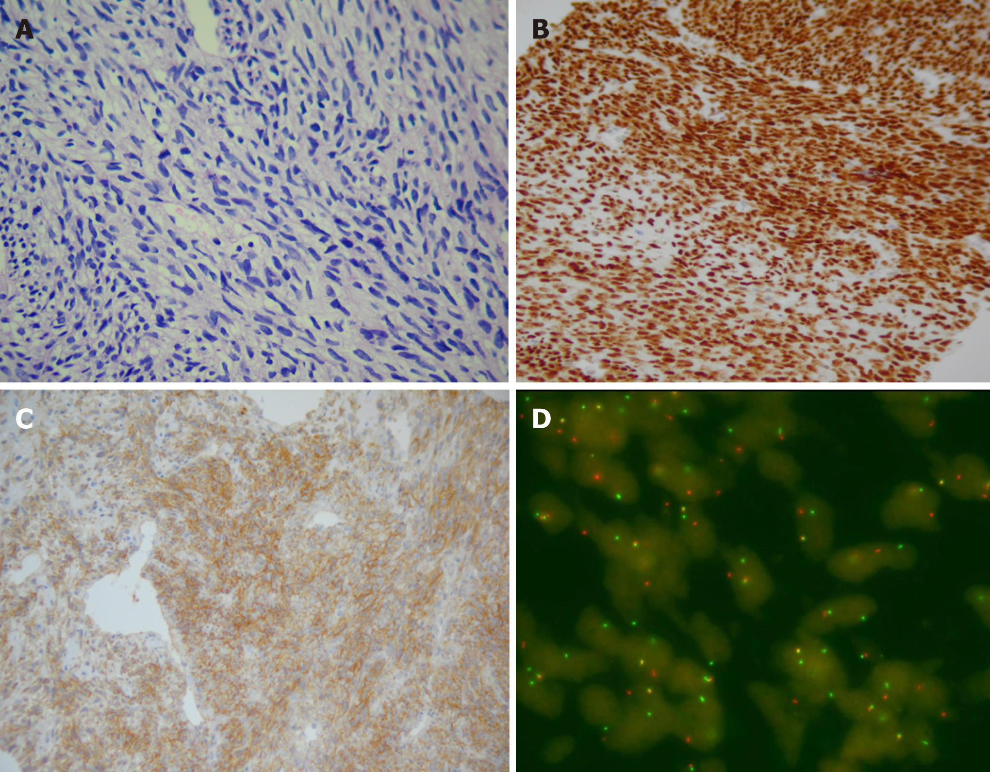
Figure 3 Pathological results of the neoplasms.
A: HE-stained image (200×) showing monotonous neoplasms formed by spindle-shaped cells; B and C: Immunohistochemical analysis demonstrated that the neoplasms expressed TLE (B, 100×) and vimentin CD99 (C, 200×); D: Genetic analysis confirmed the presence of SYT-SSX translocation using a SYT dual color break apart probe-based FISH test. About 50% of the tumor cells had abnormal yellow, red, and green break signals.
- Citation: Xu RF, He EH, Yi ZX, Lin J, Zhang YN, Qian LX. Multimodality-imaging manifestations of primary renal-allograft synovial sarcoma: First case report and literature review. World J Clin Cases 2019; 7(13): 1677-1685
- URL: https://www.wjgnet.com/2307-8960/full/v7/i13/1677.htm
- DOI: https://dx.doi.org/10.12998/wjcc.v7.i13.1677


