Copyright
©The Author(s) 2019.
World J Clin Cases. Jul 6, 2019; 7(13): 1677-1685
Published online Jul 6, 2019. doi: 10.12998/wjcc.v7.i13.1677
Published online Jul 6, 2019. doi: 10.12998/wjcc.v7.i13.1677
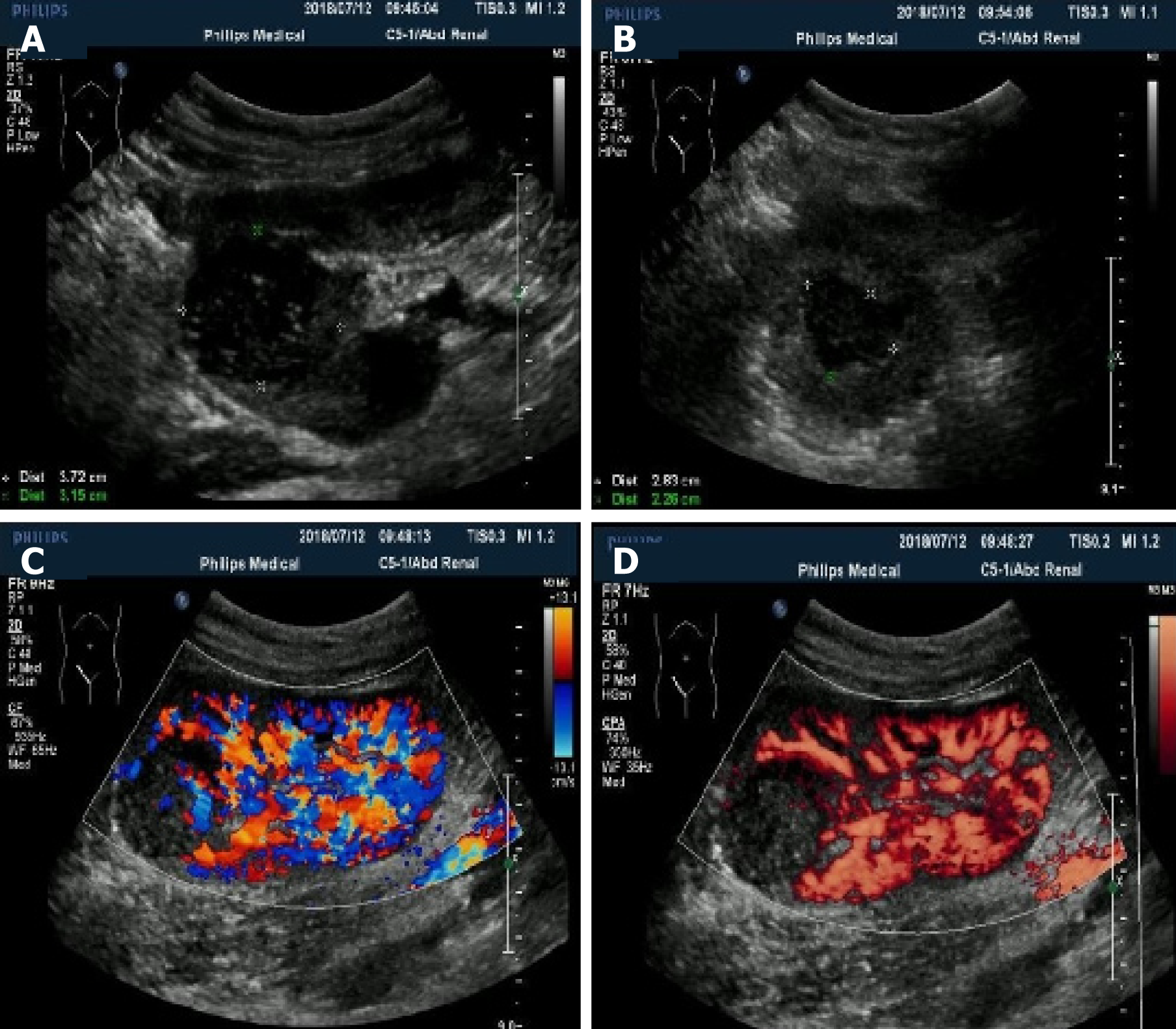
Figure 1 Ultrasonography manifestation of the neoplasms.
A and B: Conventional ultrasound showed well-defined solid hypo-echo neoplasms in the renal allograft; C and D: Color Doppler flow imaging and power Doppler imaging revealed punctiform blood flow signals inside the neoplasms.
- Citation: Xu RF, He EH, Yi ZX, Lin J, Zhang YN, Qian LX. Multimodality-imaging manifestations of primary renal-allograft synovial sarcoma: First case report and literature review. World J Clin Cases 2019; 7(13): 1677-1685
- URL: https://www.wjgnet.com/2307-8960/full/v7/i13/1677.htm
- DOI: https://dx.doi.org/10.12998/wjcc.v7.i13.1677


