Copyright
©The Author(s) 2019.
World J Clin Cases. Jul 6, 2019; 7(13): 1643-1651
Published online Jul 6, 2019. doi: 10.12998/wjcc.v7.i13.1643
Published online Jul 6, 2019. doi: 10.12998/wjcc.v7.i13.1643
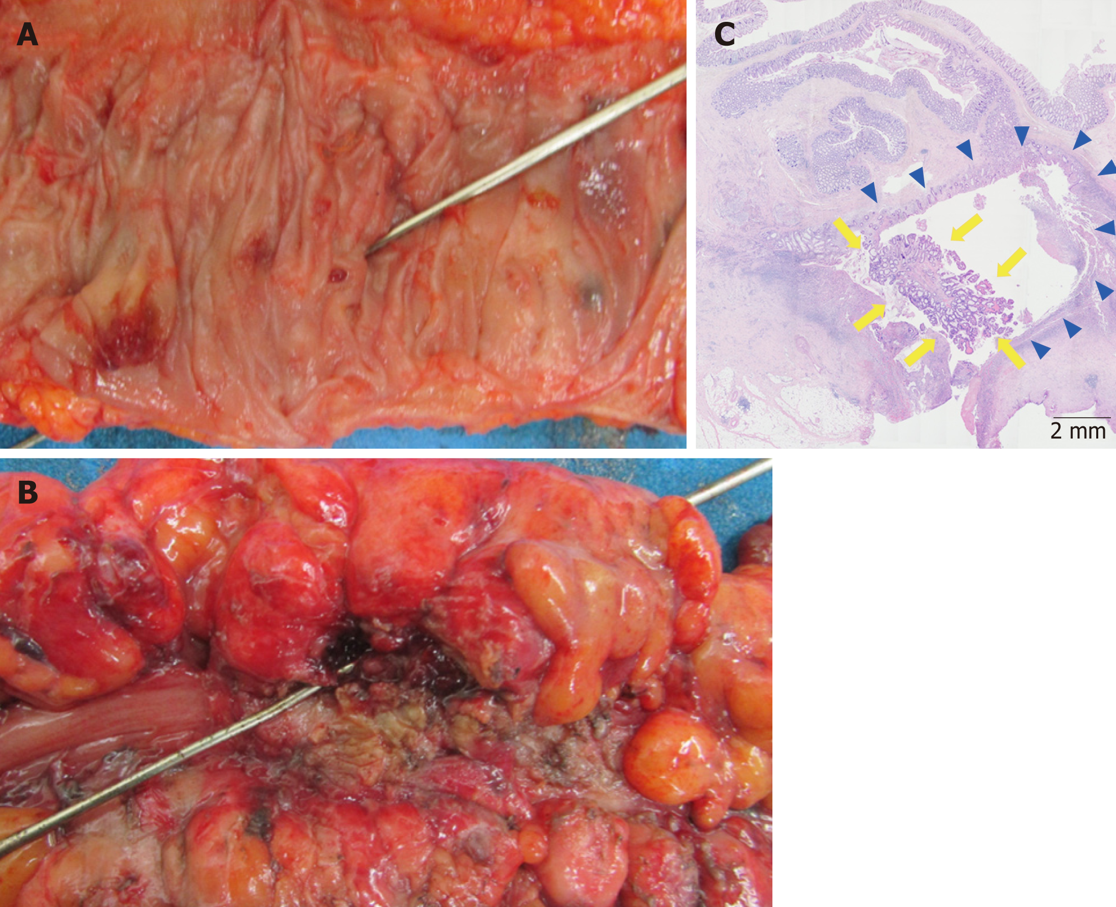
Figure 3 Histopathological examination findings.
A, B: Macroscopically, several diverticulum are visible in the colonic lumen, but tumorous lesions around the fistula are not observed; C: Histologically, there is tumor characterized by mild exophytic papillary growth (arrowheads) along the diverticular lumen (arrows). It was diagnosed as well-differentiated adenocarcinoma (HE staining, × 100).
- Citation: Kayano H, Ueda Y, Machida T, Hiraiwa S, Zakoji H, Tajiri T, Mukai M, Nomura E. Colon cancer arising from colonic diverticulum: A case report. World J Clin Cases 2019; 7(13): 1643-1651
- URL: https://www.wjgnet.com/2307-8960/full/v7/i13/1643.htm
- DOI: https://dx.doi.org/10.12998/wjcc.v7.i13.1643


