Copyright
©The Author(s) 2019.
World J Clin Cases. Jul 6, 2019; 7(13): 1643-1651
Published online Jul 6, 2019. doi: 10.12998/wjcc.v7.i13.1643
Published online Jul 6, 2019. doi: 10.12998/wjcc.v7.i13.1643
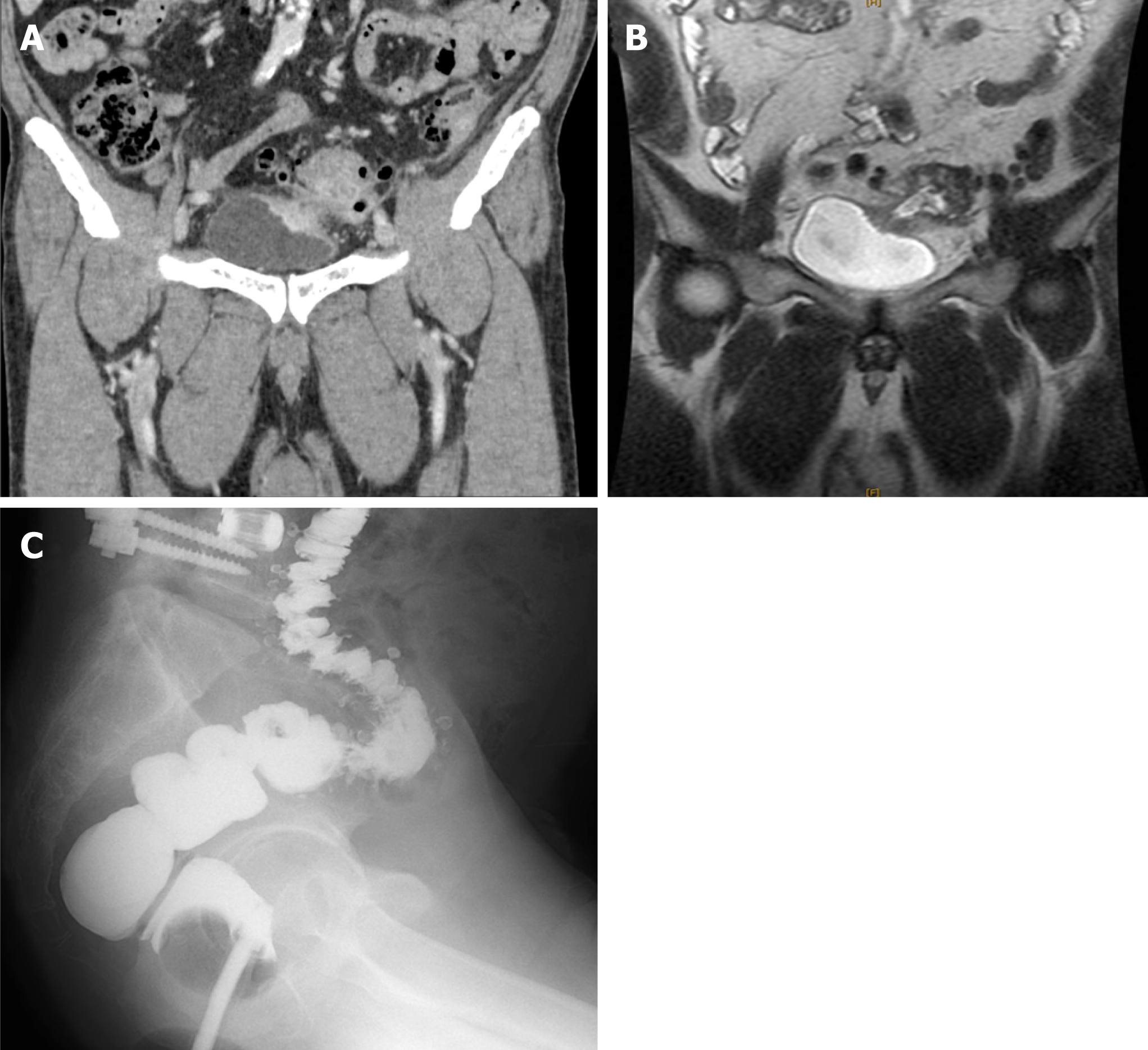
Figure 1 Preoperative computed tomography, magnetic resonance imaging, and gastrografin enema examination.
A: The inflammatory changes around the intestinal tract continue to the left bladder wall, and the bladder wall is slightly thickened. Tumor cannot be seen; B: A penetrating portion continuous with the sigmoid colon is visible on the upper left side wall of the bladder; C: Only a diverticulum is found in the sigmoid colon.
- Citation: Kayano H, Ueda Y, Machida T, Hiraiwa S, Zakoji H, Tajiri T, Mukai M, Nomura E. Colon cancer arising from colonic diverticulum: A case report. World J Clin Cases 2019; 7(13): 1643-1651
- URL: https://www.wjgnet.com/2307-8960/full/v7/i13/1643.htm
- DOI: https://dx.doi.org/10.12998/wjcc.v7.i13.1643


