Copyright
©The Author(s) 2019.
World J Clin Cases. Jun 26, 2019; 7(12): 1515-1521
Published online Jun 26, 2019. doi: 10.12998/wjcc.v7.i12.1515
Published online Jun 26, 2019. doi: 10.12998/wjcc.v7.i12.1515
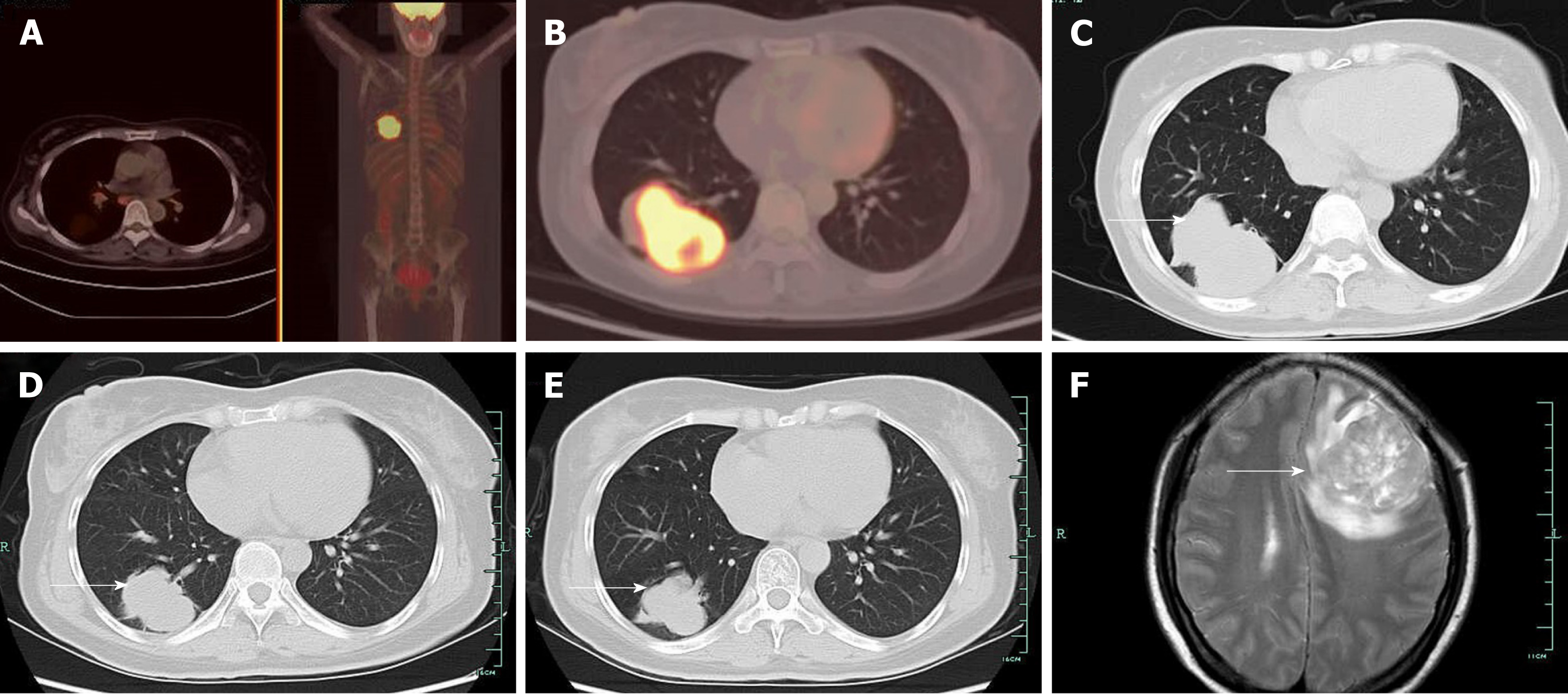
Figure 1 Case 1.
A: Positron emission tomography-computed tomography (PET-CT) was performed before lung biopsy and showed hyper-metabolic activity in pulmonary lesions and the cervix uteri; B: PET-CT image showed hyper-metabolic activity in the inferior lobe of the right lung, where lung biopsy was performed; C: CT showed a right pulmonary mass characterized by a solid region with contiguous ground-glass areas, stellate borders, and pleural puckering before tyrosine kinase inhibitor treatment; D: CT showed a right pulmonary mass after 2 mo of gefitinib therapy, which indicated partial remission of tumor; E: CT image showed a right pulmonary mass after 4 mo of gefitinib therapy; F: Magnetic resonance imaging performed on April 12, 201 indicated brain metastasis.
- Citation: Yan RL, Wang J, Zhou JY, Chen Z, Zhou JY. Female genital tract metastasis of lung adenocarcinoma with EGFR mutations: Report of two cases. World J Clin Cases 2019; 7(12): 1515-1521
- URL: https://www.wjgnet.com/2307-8960/full/v7/i12/1515.htm
- DOI: https://dx.doi.org/10.12998/wjcc.v7.i12.1515


