Copyright
©The Author(s) 2019.
World J Clin Cases. Jun 26, 2019; 7(12): 1410-1420
Published online Jun 26, 2019. doi: 10.12998/wjcc.v7.i12.1410
Published online Jun 26, 2019. doi: 10.12998/wjcc.v7.i12.1410
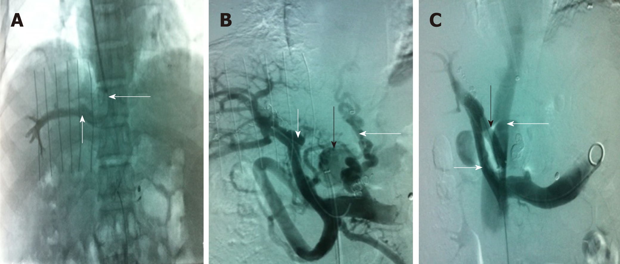
Figure 1 Transjugular intrahepatic portosystemic shunt.
A: Venography of the right hepatic vein via jugular vein catheterization showed that the sharp angles/spatial relationship between the inferior vena cava (long white arrow) and the right hepatic vein (short white arrow) were inappropriate; thus, conventional transjugular intrahepatic portosystemic shunt could not be performed; B: Puncture of the intrahepatic portal vein through the inferior vena cava via femoral vein access and venography. The venogram showed the portal vein (short white arrow), lateral branch (black arrow) and varicose vein (long white arrow); C: The shunt (black arrow) between the portal vein (short white arrow) and the inferior vena cava (long white arrow) was successfully created. Subsequently, the varicose veins and collateral vessels disappeared completely.
- Citation: Zhang Y, Liu FQ, Yue ZD, Zhao HW, Wang L, Fan ZH, He FL. Safety and efficacy of transfemoral intrahepatic portosystemic shunt for portal hypertension: A single-center retrospective study. World J Clin Cases 2019; 7(12): 1410-1420
- URL: https://www.wjgnet.com/2307-8960/full/v7/i12/1410.htm
- DOI: https://dx.doi.org/10.12998/wjcc.v7.i12.1410


